An unusual case of superior vena cava syndrome caused by the intravascular invasion of an invasive thymoma
- PMID: 24348669
- PMCID: PMC3861377
- DOI: 10.4046/trd.2013.75.5.210
An unusual case of superior vena cava syndrome caused by the intravascular invasion of an invasive thymoma
Abstract
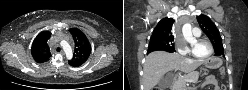
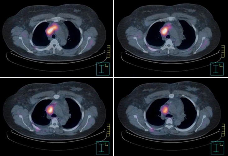
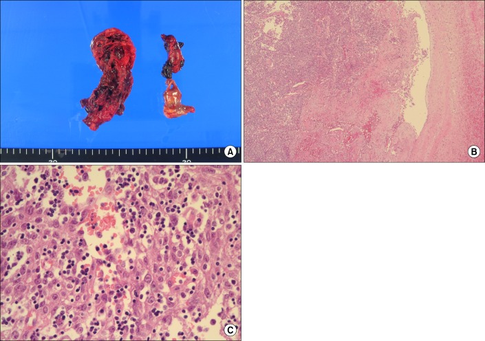
Superior vena cava syndrome (SVCS) is usually caused by extrinsic compression or invasion of the superior vena cava (SVC) by malignant tumors involving mediastinal structures. Although thymomas are well-known causes of SVCS, cases of SVCS caused by malignant thymomas protruding into adjacent vessels draining the SVC with thrombosis have been very rarely reported worldwide. We experienced a 39-year-old female patient with SVCS that developed after the direct invasion of the left brachiocephalic vein (LBCV) and SVC by an anterior mediastinal mass with a high maximum standardized uptake value on the chest computed tomography (CT) and positron emission tomography-CT. Based on these results, she underwent en bloc resection of the tumor, including removal of the involved vessels, and was eventually diagnosed as having a type B2 thymoma permeating into the LBCV and SVC. We present this case as a very rare form of SVCS caused by an invasive thymoma.
Keywords: Positron-Emission Tomography; Superior Vena Cava Syndrome; Thymoma.
Figures
Similar articles
-
An invasive thymoma extending into the superior vena cava and right atrium.Ann Transl Med. 2019 Sep;7(18):498. doi: 10.21037/atm.2019.08.59. Ann Transl Med. 2019. PMID: 31700934 Free PMC article.
-
Superior vena cava replacement combined with venovenous shunt for thymic carcinoma.Int J Surg Case Rep. 2020;72:104-107. doi: 10.1016/j.ijscr.2020.05.069. Epub 2020 Jun 6. Int J Surg Case Rep. 2020. PMID: 32534411 Free PMC article.
-
The superior vena cava syndrome caused by malignant disease. Imaging with multi-detector row CT.Eur J Radiol. 2006 Jul;59(1):93-103. doi: 10.1016/j.ejrad.2006.01.003. Epub 2006 Feb 14. Eur J Radiol. 2006. PMID: 16476534
-
Malignant Superior Vena Cava Syndrome: State of the Art.Cureus. 2022 Jan 4;14(1):e20924. doi: 10.7759/cureus.20924. eCollection 2022 Jan. Cureus. 2022. PMID: 35004083 Free PMC article. Review.
-
INTRAVASCULAR LIPOMA OF THE RIGHT BRACHIOCEPHALIC VEIN AND SUPERIOR VENA CAVA: A CASE REPORT AND LITERATURE REVIEW.Georgian Med News. 2021 Feb;(311):33-36. Georgian Med News. 2021. PMID: 33814386 Review.
Cited by
-
Invasive thymoma presenting as classic superior vena cava syndrome: a case of venous spread metastasis.BMJ Case Rep. 2016 Oct 26;2016:bcr2016217695. doi: 10.1136/bcr-2016-217695. BMJ Case Rep. 2016. PMID: 27797848 Free PMC article.
-
Thymoma Causing Bilateral Upper Extremity Deep Vein Thrombosis.Respir Med Case Rep. 2020 Apr 3;30:101049. doi: 10.1016/j.rmcr.2020.101049. eCollection 2020. Respir Med Case Rep. 2020. PMID: 32300521 Free PMC article.
-
Giant thymoma presenting as a large bilateral intrathoracic mass: A case report and a comparison between median sternotomy and hemiclamshell approach.Ann Med Surg (Lond). 2021 Sep 13;70:102859. doi: 10.1016/j.amsu.2021.102859. eCollection 2021 Oct. Ann Med Surg (Lond). 2021. PMID: 34584686 Free PMC article.
-
Giant thymoma successfully resected via hemiclamshell thoracotomy: a case report.J Thorac Dis. 2016 Aug;8(8):E677-80. doi: 10.21037/jtd.2016.06.41. J Thorac Dis. 2016. PMID: 27621898 Free PMC article.
-
Thymomas With Intravascular and Intracardiac Growth.Front Oncol. 2022 Jun 24;12:881553. doi: 10.3389/fonc.2022.881553. eCollection 2022. Front Oncol. 2022. PMID: 35814455 Free PMC article.
References
-
- Herscovici R, Szyper-Kravitz M, Altman A, Eshet Y, Nevo M, Agmon-Levin N, et al. Superior vena cava syndrome: changing etiology in the third millennium. Lupus. 2012;21:93–96. - PubMed
-
- Wan JF, Bezjak A. Superior vena cava syndrome. Emerg Med Clin North Am. 2009;27:243–255. - PubMed
-
- Yu M, Shi A, Jin B, Jiang X, Liang H, Ouyang C. Superior vena cava occlusion caused by Behcet disease. J Vasc Surg. 2012;55:1488–1491. - PubMed
-
- Airan B, Sharma R, Iyer KS, Kalia PK, Singh MK, Shrivastava S, et al. Malignant thymoma presenting as intracardiac tumor and superior vena caval obstruction. Ann Thorac Surg. 1990;50:989–991. - PubMed
-
- Terada Y, Ono N, Noguchi T, Kamakari K, Kitano M. A case of thymoma protruding into the superior vena cava through the thymic vein. Ann Thorac Surg. 2004;77:1088–1090. - PubMed
LinkOut - more resources
Full Text Sources
Other Literature Sources
Research Materials