. 2013:2013:953162.
doi: 10.1155/2013/953162. Epub 2013 Nov 20.

Effects of pretreatment with a combination of melatonin and electroacupuncture in a rat model of transient focal cerebral ischemia

Affiliations

Effects of pretreatment with a combination of melatonin and electroacupuncture in a rat model of transient focal cerebral ischemia

Lingguang Liu et al. Evid Based Complement Alternat Med. 2013.

Abstract

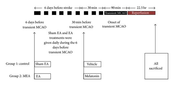
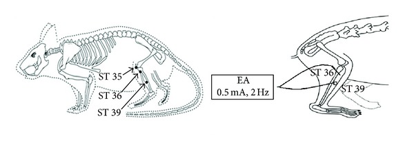
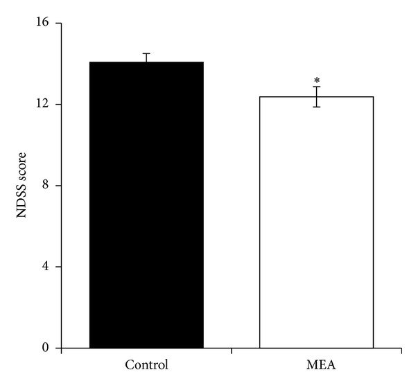
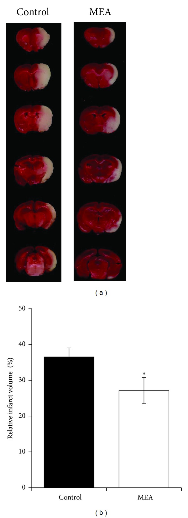
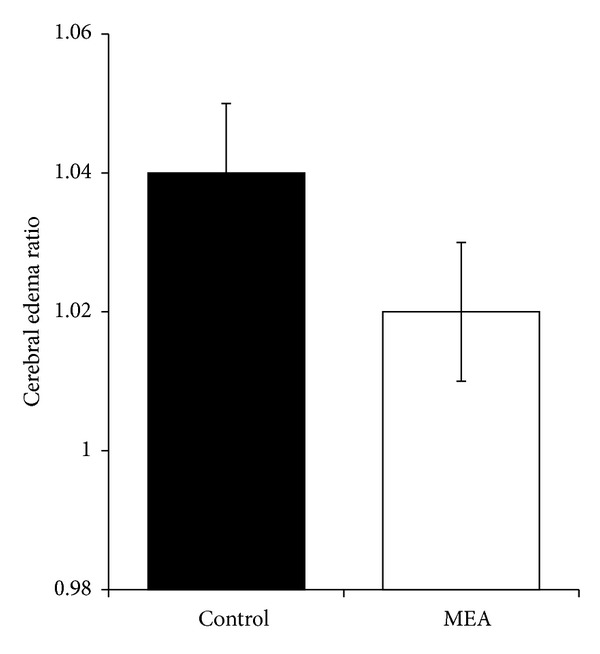
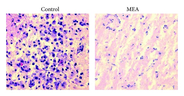
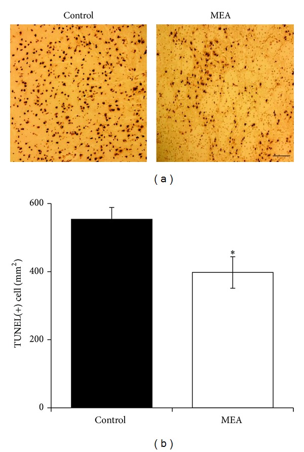
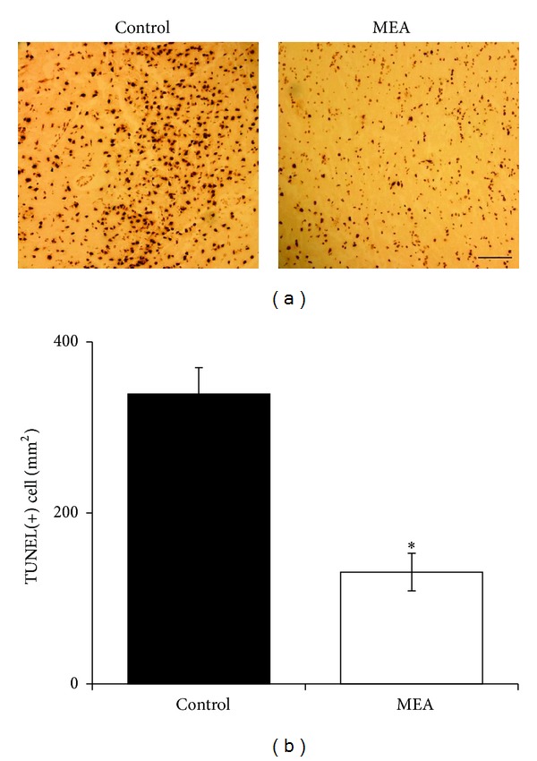
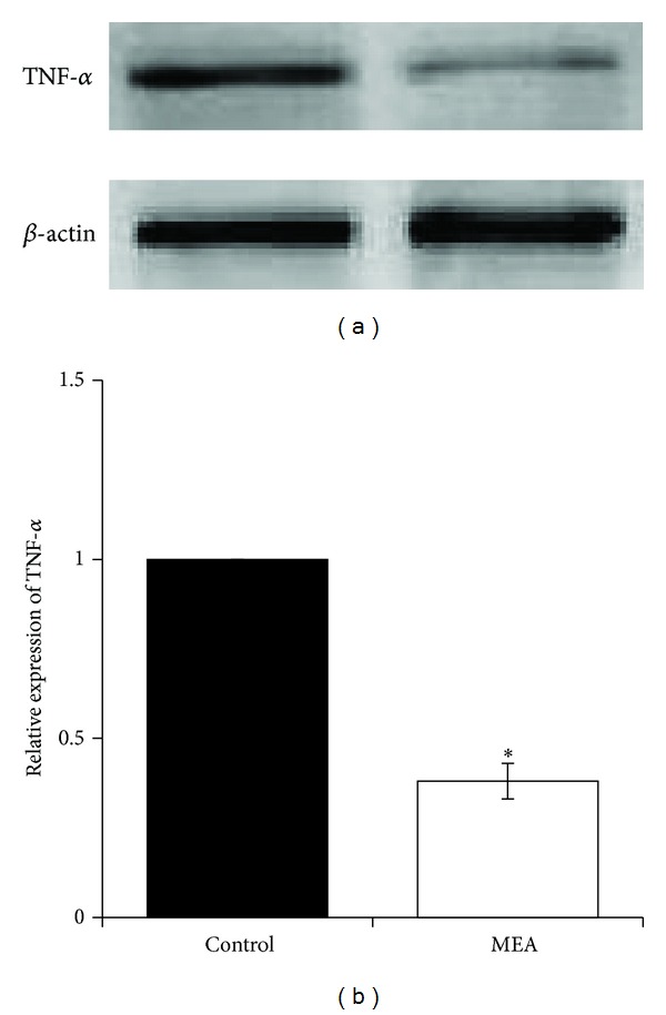
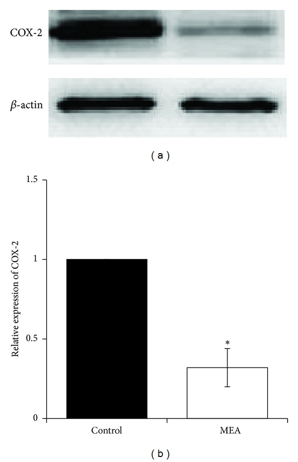
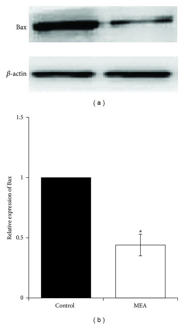
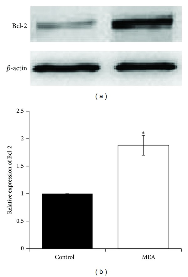
Both melatonin and electroacupuncture (EA) have been suggested to be effective treatments against stroke. However, it is unknown whether a combination of these two therapies could be beneficial against transient focal cerebral ischemia. The present study investigated the effects of pretreatment of a combination of melatonin and EA in a rat model of transient middle cerebral artery occlusion (MCAO). After pretreatment of melatonin plus EA (MEA), transient MCAO was induced for 90 minutes in male Sprague-Dawley (SD) rats. The neurological deficit score, brain infarct volume, cerebral edema ratio, neuronal inflammation, and apoptosis were evaluated 24 hours after transient MCAO. The expression of related inflammatory and apoptotic mediators in the brain was also investigated. The results showed that MEA improved neurological outcome, reduced brain infarct volume, and inhibited neuronal inflammation as well as apoptosis 24 hours after transient MCAO. The beneficial effects may derive from downregulation of proinflammatory and proapoptotic mediators and upregulation of antiapoptotic mediators. Thus, these results suggest a preventive effect of pretreatment of MEA on transient focal cerebral ischemia.


Figures

Figure 1
Figure 1
Schematic overview of the experimental procedures in a rat model of transient MCAO. Two groups are included in the experiment. The horizontal line represents time. A single i.p. injection of the vehicle and melatonin was given 30 minutes before transient MCAO in control and MEA groups, respectively. Whilst the rats were under anesthesia, sham and real EA were given daily 6 days before transient MCAO in the sham EA group and EA group, respectively. All the rats were killed 24 hours after transient MCAO for determination of brain infarct volume, cerebral edema ration and tissue processing.
Figure 2
Figure 2
Diagram of the acupoints location and acupuncture manipulation in the rat. ST 35, dubi, is located at the depression lateral to the patella ligament; ST 36, zusanli, is located at proximal one-fifth point on the line from ST 35 to the anterior side of ankle crease; ST 39, xiajuxu, is located at proximal 3 fifths point on the line from ST 35 to the anterior side of ankle crease.
Figure 3
Figure 3
Neurological deficit scoring system (NDSS) score 24 hours after transient MCAO. Data are expressed as mean ± SEM (n = 14). *P < 0.05, compared with the control group. The neurological function was significantly improved by MEA pretreatment.
Figure 4
Figure 4
Brain infarct volume measurement 24 hours after transient MCAO. (a) Digital photographs of the 2 mm thick coronal brain slices between the bregma levels +4 mm (anterior) and −6 mm (posterior) in control and MEA groups 24 hours after right-sided endovascular transient MCAO. TTC reaction showed viable brain tissue in red and infarcted brain tissue in white. (b) Quantitative analyses of brain infarct volume 24 hours after transient MCAO. The data are expressed as percentage of the contralateral hemispheric volume (mean ± SEM, in %, n = 14). *Indicates the significant difference between the control group and MEA group (P < 0.05).
Figure 5
Figure 5
Data of cerebral edema ratio 24 hours after transient MCAO. Data are expressed as mean ± SEM (n = 14). Student's t-test reveals no significant difference in cerebral edema ratio 24 hours after transient MCAO between the control and MEA group (P > 0.05).
Figure 6
Figure 6
Photomicrographs of HE-stained brain sections near bregma level 0 24 hours after right-sided transient MCAO (magnification: 400x). In the control group, neutrophil infiltration was mainly present in the infarcted cortex. Neutrophil infiltration in the ischemic cerebral cortex was suppressed by the MEA pretreatment group 24 hours after transient MCAO.
Figure 7
Figure 7
Apoptosis in the infarcted cortex of right ischemic cerebral hemisphere 24 hours after transient MCAO. (a) Representative images of TUNEL staining in the infarcted cortex of right ischemic cerebral hemisphere 24 hours after transient MCAO. The brown staining within the nuclei reveals TUNEL-positive cells. (b) Quantitative analysis of TUNEL-positive cells. Data are expressed as means ± SEM (n = 5). *P < 0.05, compared with the control group. The number of TUNEL-positive cells was significantly decreased by MEA pretreatment. Scale bar = 100 μm.
Figure 8
Figure 8
Apoptosis in the penumbra of right ischemic cerebral hemisphere 24 hours after transient MCAO. (a) Representative images of TUNEL staining in the penumbra of right ischemic cerebral hemisphere 24 hours after transient MCAO. The brown staining within the nuclei reveals TUNEL-positive cells. (b) Quantitative analysis of TUNEL-positive cells. Data are expressed as means ± SEM (n = 5). *P < 0.01, compared with the control group. The number of TUNEL-positive cells was significantly decreased by MEA pretreatment. Scale bar = 100 μm.
Figure 9
Figure 9
Protein expression of TNF-α 24 hours after transient MCAO in different groups. (a) Representative immunoblots of TNF-α in the right cerebral hemisphere of different groups 24 hours after transient MCAO. (b) Semiquantitative analysis of protein expression of TNF-α. Data are expressed as means ± SEM (n = 3). *P < 0.01, compared with the control group. The relative expression of TNF-α was significantly inhibited by MEA pretreatment.
Figure 10
Figure 10
Protein expression of COX-2 24 hours after transient MCAO in different groups. (a) Representative immunoblots of COX-2 in the right cerebral hemisphere of different groups 24 hours after transient MCAO. (b) Semiquantitative analysis of protein expression of COX-2. Data are expressed as means ± SEM (n = 3). *P < 0.01, compared with the control group. The relative expression of COX-2 was significantly decreased by MEA pretreatment.
Figure 11
Figure 11
Protein expression of Bax 24 hours after transient MCAO in different groups. (a) Representative immunoblots of Bax in the right cerebral hemisphere of different groups 24 hours after transient MCAO. (b) Semiquantitative analysis of protein expression of Bax. Data are expressed as means ± SEM (n = 3). *P < 0.05, compared with the control group. The relative expression of Bax was significantly decreased by MEA pretreatment.
Figure 12
Figure 12
Protein expression of Bcl-2 24 hours after transient MCAO in different groups. (a) Representative immunoblots of Bcl-2 in the right cerebral hemisphere of different groups 24 hours after transient MCAO. (b) Semiquantitative analysis of protein expression of Bcl-2. Data are expressed as means ± SEM (n = 3). *P < 0.01, compared with the control group. The relative expression of Bcl-2 was significantly increased by MEA pretreatment.

Similar articles

Cited by

References

    1. Hacke W, Kaste M, Bluhmki E, et al. Thrombolysis with alteplase 3 to 4.5 hours after acute ischemic stroke. The New England Journal of Medicine. 2008;359(13):1317–1329. - PubMed
    1. Cheng YD, Al-Khoury L, Zivin JA. Neuroprotection for ischemic stroke: two decades of success and failure. NeuroRx. 2004;1(1):36–45. - PMC - PubMed
    1. Faden AI, Stoica B. Neuroprotection: challenges and opportunities. Archives of Neurology. 2007;64(6):794–800. - PubMed
    1. Zhang L, Zhang ZG, Chopp M. The neurovascular unit and combination treatment strategies for stroke. Trends in Pharmacological Sciences. 2012;33(8):415–422. - PMC - PubMed
    1. Adeoye O, Hornung R, Khatri P, Kleindorfer D. Recombinant tissue-type plasminogen activator use for ischemic stroke in the united states: a doubling of treatment rates over the course of 5 years. Stroke. 2011;42(7):1952–1955. - PMC - PubMed

LinkOut - more resources