. 2014 Jan;7(1):31-38.
doi: 10.3892/etm.2013.1391. Epub 2013 Nov 7.

Null anticarcinogenic effect of silymarin on diethylnitrosamine-induced hepatocarcinogenesis in rats

Affiliations

Null anticarcinogenic effect of silymarin on diethylnitrosamine-induced hepatocarcinogenesis in rats

Ryu Imamoto et al. Exp Ther Med. 2014 Jan.

Abstract

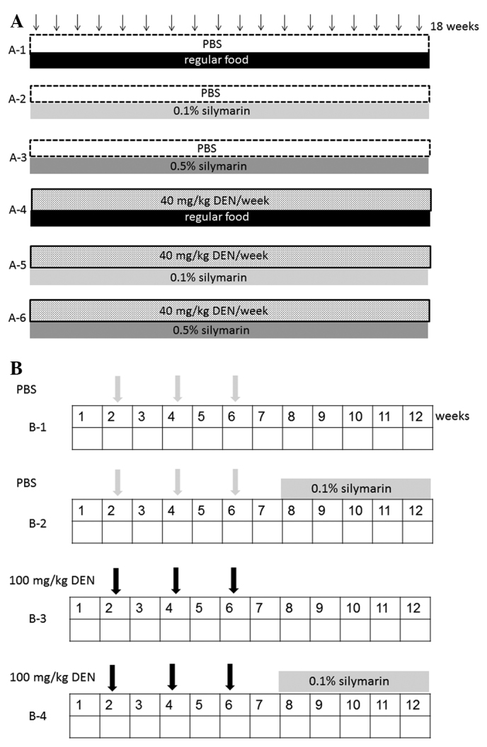
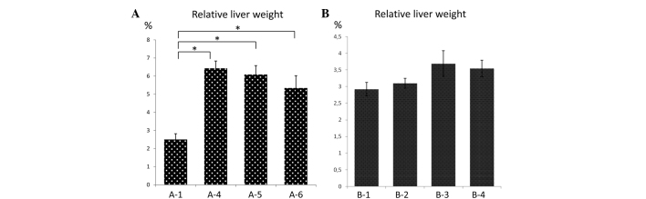
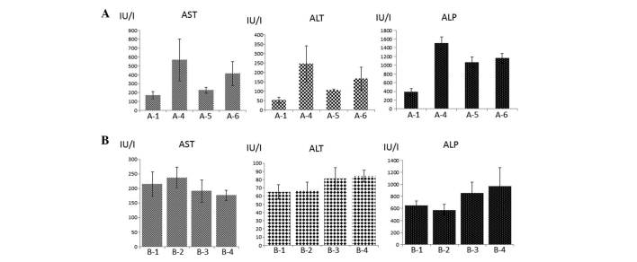
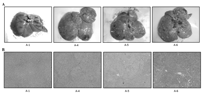
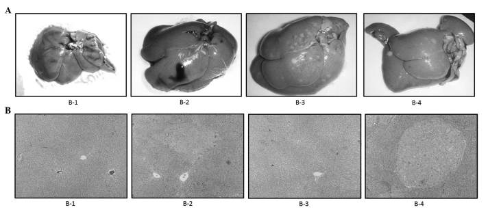
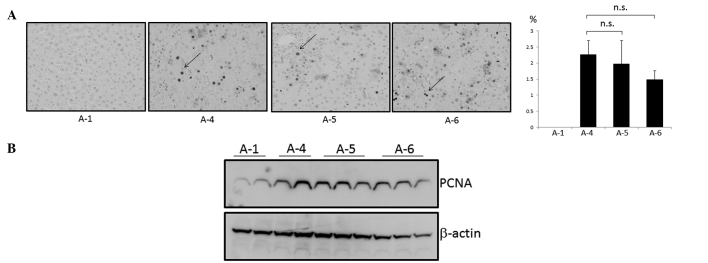
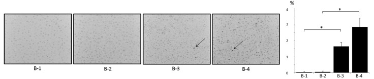
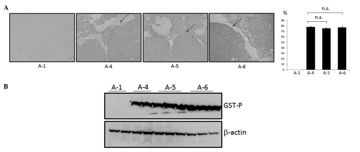
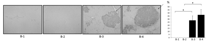
The aim of this study was to investigate the anticarcinogenic effects of silymarin in diethylnitrosamine (DEN)-induced hepatocarcinogenic rat models. Severe and mild models of hepatocellular carcinoma (HCC) were generated by the intraperitoneal administration of 40 mg/kg DEN once a week for 18 weeks and 100 mg/kg DEN every 2 weeks for 6 weeks in male Wistar rats, respectively. In the severe and mild models of HCC, the rats were treated with 0.1 and 0.5% silymarin for 18 weeks and with 0.1% silymarin for 5 weeks, respectively. Serum transaminase levels were not significantly decreased by the silymarin treatment in either model. Macroscopic and microscopic features indicated that the silymarin-containing formulations did not significantly inhibit the hepatic tumor formation induced by DEN. Furthermore, immunohistochemical and western blot analyses demonstrated that the expression levels of proliferating cell nuclear antigen and glutathione S-transferase P, which are hepatocarcinogenic markers, were not significantly modified by the silymarin treatment. These results indicate that silymarin may not be considered as a candidate agent against hepatocarcinogenesis.

Keywords: diethylnitrosamine; hepatocellular carcinoma; silymarin.


Figures

Figure 1
Figure 1
Experimental schedules. (A) In the severe hepatocellular carcinoma (HCC) model (model A), male Wistar rats were divided into six groups. The animals were intraperitoneally injected with 300 μl phosphate-buffered saline (PBS; groups A-1, A-2 and A-3; n=4) or diethylnitrosamine (DEN; 40 mg/kg body weight) dissolved in PBS weekly for 18 weeks (groups A-4, A-5 and A-6; n=4). The rats were fed with 0.1% silymarin (groups A-2 and A-5) and 0.5% silymarin (groups A-3 and A-6) in powder form for 18 weeks. One week subsequent to the 18-week treatments, the animals were sacrificed.(B) In the mild HCC model (model B), male Wistar rats were randomized and divided into four groups. The animals were intraperitoneally injected with 300 μl PBS (groups B-1 and B-2; n=8) or DEN (100 mg/kg body weight) dissolved in PBS (groups B-3 and B-4; n=8) at 15-day intervals on experimental weeks 2, 4 and 6. In groups B-2 and B-4, the rats were fed with 0.1% silymarin in powder form during experimental weeks 8 to 12. One week subsequent to the final treatments, the animals were sacrificed.
Figure 2
Figure 2
Relative liver weight (liver weight/body weight). Means of relative liver weight in (A) severe model groups A-1, A-4, A-5 and A-6 and (B) mild model groups B-1, B-2, B-3 and B-4. *P<0.05.
Figure 3
Figure 3
Serum transaminase and alkaline phosphatase (ALP) levels. Means of serum alanine aminotransferase (ALT), aspartate aminotransferase (AST) and ALP levels in groups (A) A-1, A-4, A-5 and A-6 and (B) B-1, B-2, B-3 and B-4.
Figure 4
Figure 4
Representative (A) macroscopic and (B) microscopic features of the livers of model A rats. (A) In control rats treated with phosphate-buffered saline (PBS) without diethylnitrosamine (DEN), no tumors were observed (A-1, A-2 and A-3; data not shown for A-2 and A-3). Multiple white nodules were macroscopically observed following DEN administration, irrespective of silymarin treatment (A-4, A-5 and A-6). (B) In control rats treated with PBS without DEN, the liver histology showed a normal appearance (A-1, A-2 and A-3; data not shown for A-2 and A-3). In the histological analysis, white nodules induced by DEN were demonstrated to be hepatocellular carcinoma (HCC; A-4). The HCC area was not significantly modified by silymarin treatment (A-5 and A-6); original magnification, ×100.
Figure 5
Figure 5
Representative (A) macroscopic and (B) microscopic features of the livers of model B rats. (A) Several white nodules were macroscopically observed following diethylnitrosamine (DEN) treatment (B-3). The gross appearance of the liver treated with DEN and 0.1% silymarin was mostly identical to that of the liver treated with DEN alone (B-4). (B) In the histological analysis, hyperplastic nodules were developed following treatment with DEN (B-3), which was not significantly modified by the silymarin treatment (B-4); original magnification, ×100.
Figure 6
Figure 6
Expression of proliferating cell nuclear antigen (PCNA) in the livers of model A rats. Expression levels were evaluated using (A) immunohistochemical and (B) western blot analyses. (A) Representative liver tissues immunostained with anti-PCNA antibody in the control (A-1), diethylnitrosamine (DEN; A-4), DEN with 0.1% silymarin (A-5) and DEN with 0.5% silymarin (A-6) groups (top left panel). Arrows indicate the representative PCNA-positive cells. Percentages of PCNA-positive cells in A-1, A-4, A-5 and A-6 were 0, 2.3, 2.0 and 1.5%, respectively (top right panel); original magnification, ×400. (B) Representative liver samples from groups A-1, A-4, A-5 and A-6 were probed with anti-PCNA antibody (top lane). The membrane was reprobed with anti-β-actin antibody (bottom lane). n.s., not significant.
Figure 7
Figure 7
Expression of proliferating cell nuclear antigenin (PCNA) in the livers of model B rats. Representative liver tissues immunostained with anti-PCNA antibody in the control (B-1), 0.1% silymarin (B-2), diethylnitrosamine (DEN; B-3) and DEN with 0.1% silymarin (B-4) groups (left panel). Arrows indicate the representative PCNA-positive cells. Percentages of PCNA-positive cells in B-1, B-2, B-3 and B-4 were 0, 0, 1.7 and 2.9%, respectively (right panel); original magnification, ×400. *P<0.05.
Figure 8
Figure 8
Expression of glutathione S-transferase P (GST P) in the livers of model A rats. Expression levels were evaluated using (A) immunohistochemical and (B) western blot analyses. (A) Representative liver tissues immunostained with anti-GST P antibody in the control (A-1), diethylnitrosamine (DEN; A-4), DEN with 0.1% silymarin (A-5) and DEN with 0.5% silymarin (A-6) groups (top left panel). Arrows indicate the representative GST P-positive area. Percentages of GST P-positive areas in A-1, A-4, A-5 and A-6 were 0, 78.2, 75.5 and 77.4%, respectively (top right panel); original magnification, ×100. (B) Representative liver samples from groups A-1, A-4, A-5 and A-6 were probed with anti-GST P antibody (top lane). The membrane was reprobed with anti-β-actin antibody (bottom lane). n.s., not significant.
Figure 9
Figure 9
Expression of glutathione S-transferase P (GST P) in the livers of model B rats. Representative liver tissues immunostained with anti-GST P antibody in the control (B-1), 0.1% silymarin (B-2), diethylnitrosamine (DEN; B-3) and DEN with 0.1% silymarin (B-4) groups (left panel). Arrows indicate the representative GST P-positive area. Percentages of GST P-positive areas in B-1, B-2, B-3 and B-4 were 0, 0, 28.0 and 36.8%, respectively (right panel); original magnification, ×100. *P<0.05.

References

    1. Bosch FX, Ribes J, Borràs J. Epidemiology of primary liver cancer. Semin Liver Dis. 1999;19:271–285. - PubMed
    1. El-Serag HB, Rudolph KL. Hepatocellular carcinoma: epidemiology and molecular carcinogenesis. Gastroenterology. 2007;132:2557–2576. - PubMed
    1. Farazi PA, DePinho RA. Hepatocellular carcinoma pathogenesis: from genes to environment. Nat Rev Cancer. 2006;6:674–687. - PubMed
    1. Makuuchi M, Kokudo N, Arii S, et al. Development of evidence-based clinical guidelines for the diagnosis and treatment of hepatocellular carcinoma in Japan. Hepatol Res. 2008;38:37–51. - PubMed
    1. Avila MA, Berasain C, Sangro B, Prieto J. New therapies for hepatocellular carcinoma. Oncogene. 2006;25:3866–3884. - PubMed

LinkOut - more resources