

Giant epidermal cyst in the posterior neck developing over 40 years: A case report
- PMID: 24348807
- PMCID: PMC3861182
- DOI: 10.3892/etm.2013.1383
Giant epidermal cyst in the posterior neck developing over 40 years: A case report
Abstract
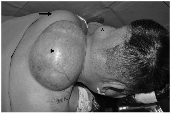
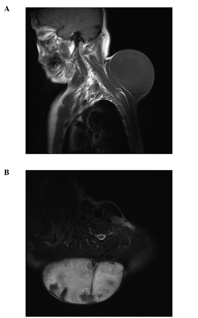
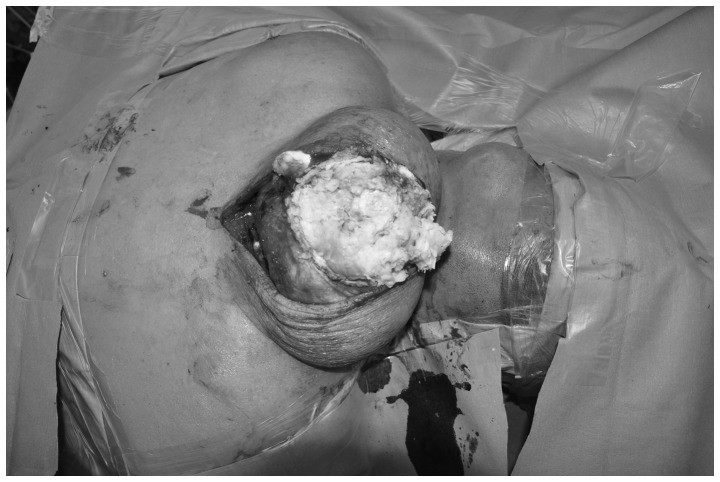
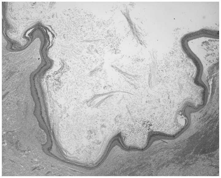
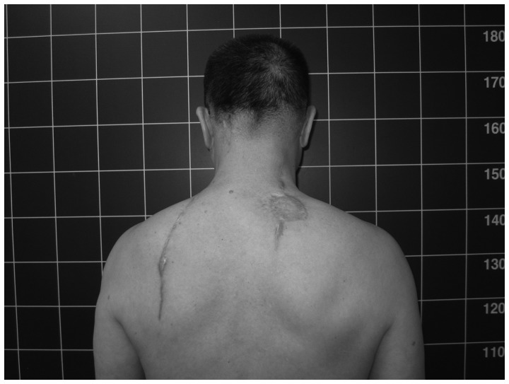
Conventional epidermal cysts are generally small, slow-growing, non-tender, dome-shaped lesions. An epidermal cyst is usually asymptomatic until it is infected or enlarged to the extent that it causes damage to adjacent anatomical structures. However, few cases of giant epidermal cysts in the neck have been reported. The present case reports a giant epidermal cyst in the posterior neck, which grew to an extremely large size for >40 years without inflammation or rupture, and was misdiagnosed as a large soft tissue neoplasm. The patient exhibited depression and developed social anxiety due to the negative cosmetic consequences of the large mass. The patient underwent excision of the mass. At the follow-up examination two years postoperatively, there were no local recurrence and the psychiatric symptoms of the patient were completely resolved. To the best of our knowledge, a giant epidermal cyst growing for >40 years has not previously been reported.
Keywords: cosmetic problem; giant epidermal cyst; posterior neck; psychiatric symptom.
Figures
References
-
- Polychronidis A, Perente S, Botaitis S, Sivridis E, Simopoulos C. Giant multilocular epidermoid cyst on the left buttock. Dermatol Surg. 2005;31:1323–1324. - PubMed
-
- Golshan Momeni M, Anavim A, Lin F, Tehranzadeh J. Giant epidermal inclusion cyst of buttock. Skeletal Radiol. 2006;35:864–866. - PubMed
-
- Kim C, Park MC, Seo SJ, Yoo YM, Jang YJ, Lee IJ. Giant epidermoid cyst of the posterior neck. J Craniofac Surg. 2011;22:1142–1144. - PubMed
-
- Sau P, Graham JH, Helwig EB. Proliferating epithelial cysts. Clinicopathological analysis of 96 cases. J Cutan Pathol. 1995;22:394–406. - PubMed
LinkOut - more resources
Full Text Sources
Other Literature Sources