

Atypical imaging observations of branchial cleft cysts
- PMID: 24348852
- PMCID: PMC3861601
- DOI: 10.3892/ol.2013.1656
Atypical imaging observations of branchial cleft cysts
Abstract
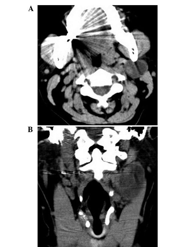
The aim of the present study was to assess the atypical imaging manifestations of branchial cleft cysts (BCCs) confirmed by pathology. Computerized tomography (CT) or magnetic resonance imaging (MRI) of 17 BCC cases were reviewed. The imaging features, including laterality, location, border, attenuation and internal architecture, were evaluated. All 17 cases were second BCCs, including 5 cases of Bailey type I classification cysts and 12 cases of type II classification cysts. The atypical imaging features included signal and morphological abnormalities. The abnormal signal intensities were caused by intracapsular bleeding (n=2) or solidification of cystic fluid (n=2). Intracystic hemorrhaging revealed homogeneous hyperintensity on T1-weighted image (T1WI) and T2-weighted image (T2WI). Solidification of cystic fluid revealed slightly homogeneous hyperintensity compared with muscle on T1WI and homogeneous hypointensity on T2WI without enhancement. The aberrant morphology mainly presented as thickening of the cystic wall (n=13). Thickened walls of BCCs with ill- (n=5) or well- (n=8) defined borders were observed in 13 patients. In 3 patients, significant enhancement was identified following intravenous gadolinium administration (n=4). When with atypical CT or MRI features are presented, the typical location of BCCs can help in the diagnosis, as it is located at the lateral portion of the neck adjacent to the anterior border of the mandibular angle or sternocleidomastoid muscle. The atypical observations, including variable signals, imply that the cystic content has changed. Thickened walls indicate inflammation or cancerous tendency and patients with ill-defined margins, vascular involvement or lymphadenopathy atelectasis indicate malignant conversion.
Keywords: branchial cleft cysts; computerized tomography; magnetic resonance imaging; neck.
Figures


References
-
- Koeller KK, Alamo L, Adair CF, Smirniotopoulos JG. Congential cystic masses of the neck: radiologic-pathologic correlation. Radiographics. 1999;19:121–146. quiz 152–153. - PubMed
-
- Bailey H. The clinical aspects of branchial cysts. Br J Surg. 1933;10:173–182.
-
- Daoud FS. Branchial cyst: an often forgotten diagnosis. Asian J Surg. 2005;28:174–178. - PubMed
LinkOut - more resources
Full Text Sources
Other Literature Sources