

Sialylation and muscle performance: sialic acid is a marker of muscle ageing
- PMID: 24349002
- PMCID: PMC3859654
- DOI: 10.1371/journal.pone.0080520
Sialylation and muscle performance: sialic acid is a marker of muscle ageing
Abstract
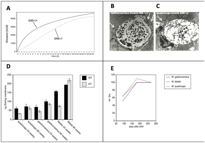
Sialic acids (Sia) are widely expressed as terminal monosaccharides on eukaryotic glycoconjugates. They are involved in many cellular functions, such as cell-cell interaction and signal recognition. The key enzyme of sialic acid biosynthesis is the bifunctional UDP-N-acetylglucosamine-2-epimerase/N-acetylmannosamine kinase (GNE), which catalyses the first two steps of Sia biosynthesis in the cytosol. In this study we analysed sialylation of muscles in wild type (C57Bl/6 GNE (+/+)) and heterozygous GNE-deficient (C57Bl/6 GNE (+/-)) mice. We measured a significantly lower performance in the initial weeks of a treadmill exercise in C57Bl/6 GNE (+/-) mice compared to wild type C57Bl/6 GNE (+/+) animals. Membrane bound Sia of C57Bl/6 GNE (+/-) mice were reduced by 33-53% at week 24 and by 12-15% at week 80 in comparison to C57Bl/6 GNE (+/+) mice. Interestingly, membrane bound Sia concentration increased with age of the mice by 16-46% in C57Bl/6 GNE (+/+), but by 87-207% in C57Bl/6 GNE (+/-). Furthermore we could identify specific morphological changes in aged muscles. Here we propose that increased Sia concentrations in muscles are a characteristic feature of ageing and could be used as a marker for age-related changes in muscle.
Conflict of interest statement
Figures
Similar articles
-
Reduced sialylation status in UDP-N-acetylglucosamine-2-epimerase/N-acetylmannosamine kinase (GNE)-deficient mice.Glycoconj J. 2007 Apr;24(2-3):125-30. doi: 10.1007/s10719-006-9019-7. Epub 2007 Jan 19. Glycoconj J. 2007. PMID: 17235685
-
Lessons from GNE-deficient embryonic stem cells: sialic acid biosynthesis is involved in proliferation and gene expression.Glycobiology. 2010 Jan;20(1):107-17. doi: 10.1093/glycob/cwp153. Epub 2009 Sep 30. Glycobiology. 2010. PMID: 19797319
-
Development of Assays to Measure GNE Gene Potency and Gene Replacement in Skeletal Muscle.J Neuromuscul Dis. 2023;10(5):797-812. doi: 10.3233/JND-221596. J Neuromuscul Dis. 2023. PMID: 37458043 Free PMC article.
-
Regulation and pathophysiological implications of UDP-GlcNAc 2-epimerase/ManNAc kinase (GNE) as the key enzyme of sialic acid biosynthesis.Biol Chem. 2009 Jul;390(7):591-9. doi: 10.1515/BC.2009.073. Biol Chem. 2009. PMID: 19426133 Review.
-
Beyond glycosylation: sialic acid precursors act as signaling molecules and are involved in cellular control of differentiation of PC12 cells.Biol Chem. 2009 Jul;390(7):575-9. doi: 10.1515/BC.2009.058. Biol Chem. 2009. PMID: 19361277 Review.
Cited by
-
Upregulation of Hallmark Muscle Genes Protects GneM743T/M743T Mutated Knock-In Mice From Kidney and Muscle Phenotype.J Neuromuscul Dis. 2020;7(2):119-136. doi: 10.3233/JND-190461. J Neuromuscul Dis. 2020. PMID: 31985472 Free PMC article.
-
Methylome-proteome integration after late-life voluntary exercise training reveals regulation and target information for improved skeletal muscle health.J Physiol. 2025 Jan;603(1):211-237. doi: 10.1113/JP286681. Epub 2024 Jul 26. J Physiol. 2025. PMID: 39058663 Free PMC article.
-
Insights into the role of sialylation in cancer progression and metastasis.Br J Cancer. 2021 Jan;124(1):76-90. doi: 10.1038/s41416-020-01126-7. Epub 2020 Nov 4. Br J Cancer. 2021. PMID: 33144696 Free PMC article. Review.
-
Risk factors and postnatal biomarkers for acute placental inflammatory lesions and intrauterine infections in preterm infants.Eur J Pediatr. 2022 Sep;181(9):3429-3438. doi: 10.1007/s00431-022-04545-1. Epub 2022 Jul 14. Eur J Pediatr. 2022. PMID: 35831682 Free PMC article.
-
Metabolomics Analysis of Skeletal Muscles from FKRP-Deficient Mice Indicates Improvement After Gene Replacement Therapy.Sci Rep. 2019 Jul 11;9(1):10070. doi: 10.1038/s41598-019-46431-1. Sci Rep. 2019. PMID: 31296900 Free PMC article.
References
-
- Hinderlich S, Stäsche R, Zeitler R, Reutter W (1997) A bifunctional enzyme catalyzes the first two steps in N-acetylneuraminic acid biosynthesis of rat liver. Purification and characterization of UDP-N-acetylglucosamine 2-epimerase/N-acetylmannosamine kinase. J Biol Chem 272: 24313–24318. - PubMed
-
- Stäsche R, Hinderlich S, Weise C, Effertz K, Lucka L, et al. (1997) A bifunctional enzyme catalyzes the first two steps in N-acetylneuraminic acid biosynthesis of rat liver. Molecular cloning and functional expression of UDP-N-acetyl-glucosamine 2-epimerase/N-acetylmannosamine kinase. J Biol Chem 272: 24319–24. - PubMed
-
- Argov Z, Yarom R (1984) “Rimmed vacuole myopathy” sparing the quadriceps. A unique disorder in Iranian Jews. J Neurol Sci 64: 33–43. - PubMed
MeSH terms
Substances
LinkOut - more resources
Full Text Sources
Other Literature Sources
Medical
Molecular Biology Databases
