. 2013 Dec 9;8(12):e81222.
doi: 10.1371/journal.pone.0081222. eCollection 2013.

Nifedipine treatment reduces resting calcium concentration, oxidative and apoptotic gene expression, and improves muscle function in dystrophic mdx mice

Affiliations

Nifedipine treatment reduces resting calcium concentration, oxidative and apoptotic gene expression, and improves muscle function in dystrophic mdx mice

Francisco Altamirano et al. PLoS One. .

Abstract

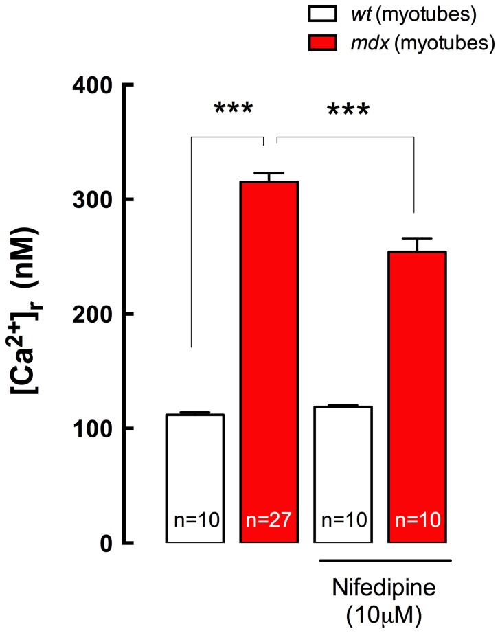
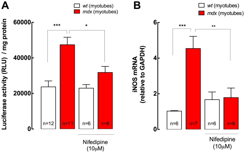
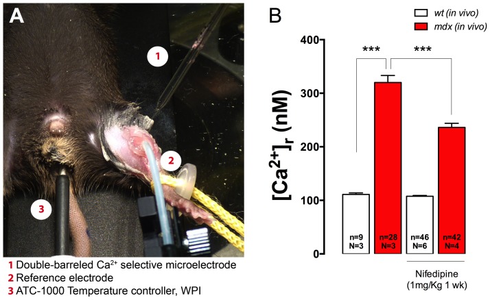
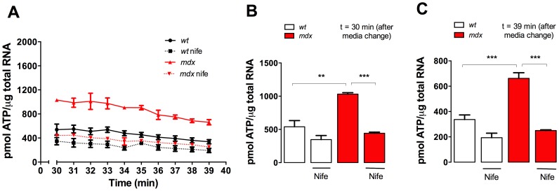
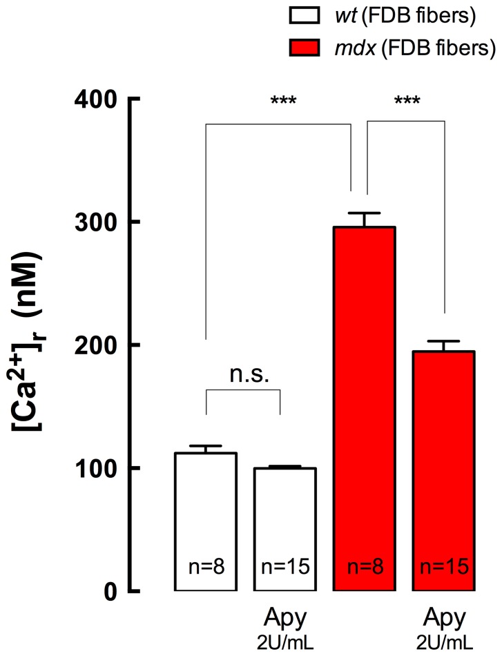
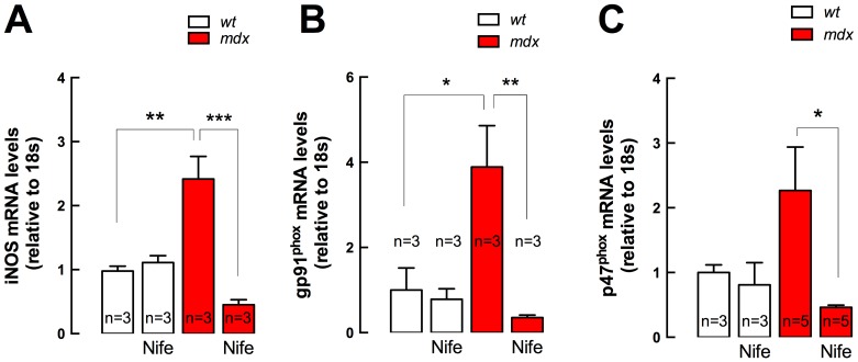
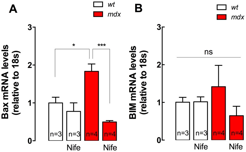
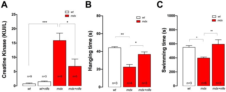
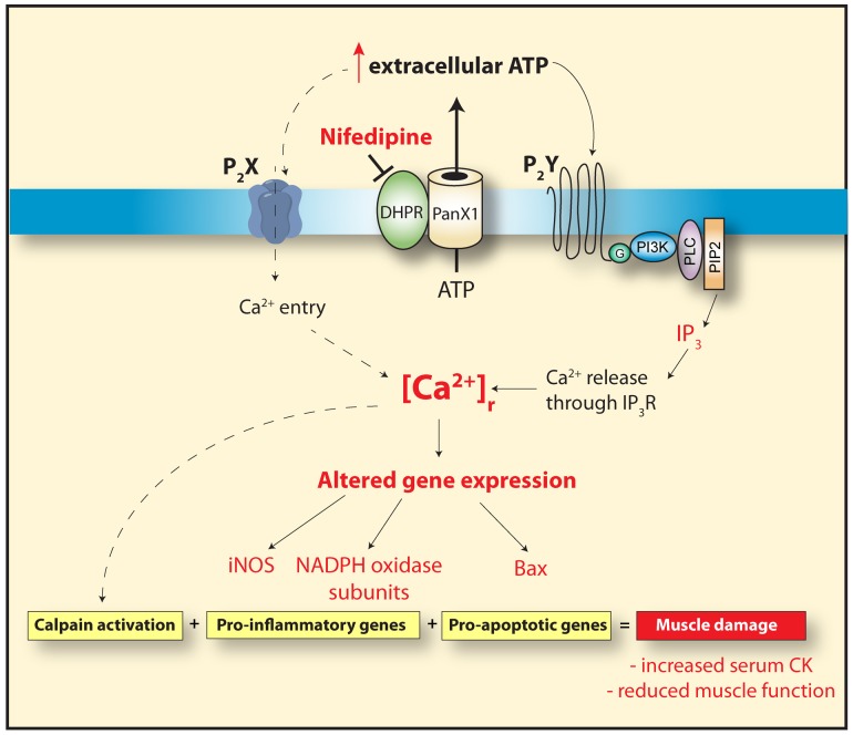
Duchenne Muscular Dystrophy (DMD) is a recessive X-linked genetic disease, caused by mutations in the gene encoding dystrophin. DMD is characterized in humans and in mdx mice by a severe and progressive destruction of muscle fibers, inflammation, oxidative/nitrosative stress, and cell death. In mdx muscle fibers, we have shown that basal ATP release is increased and that extracellular ATP stimulation is pro-apoptotic. In normal fibers, depolarization-induced ATP release is blocked by nifedipine, leading us to study the potential therapeutic effect of nifedipine in mdx muscles and its relation with extracellular ATP signaling. Acute exposure to nifedipine (10 µM) decreased [Ca(2+)]r, NF-κB activity and iNOS expression in mdx myotubes. In addition, 6-week-old mdx mice were treated with daily intraperitoneal injections of nifedipine, 1 mg/Kg for 1 week. This treatment lowered the [Ca(2+)]r measured in vivo in the mdx vastus lateralis. We demonstrated that extracellular ATP levels were higher in adult mdx flexor digitorum brevis (FDB) fibers and can be significantly reduced after 1 week of treatment with nifedipine. Interestingly, acute treatment of mdx FDB fibers with apyrase, an enzyme that completely degrades extracellular ATP to AMP, reduced [Ca(2+)]r to a similar extent as was seen in FDB fibers after 1-week of nifedipine treatment. Moreover, we demonstrated that nifedipine treatment reduced mRNA levels of pro-oxidative/nitrosative (iNOS and gp91(phox)/p47(phox) NOX2 subunits) and pro-apoptotic (Bax) genes in mdx diaphragm muscles and lowered serum creatine kinase (CK) levels. In addition, nifedipine treatment increased muscle strength assessed by the inverted grip-hanging test and exercise tolerance measured with forced swimming test in mdx mice. We hypothesize that nifedipine reduces basal ATP release, thereby decreasing purinergic receptor activation, which in turn reduces [Ca(2+)]r in mdx skeletal muscle cells. The results in this work open new perspectives towards possible targets for pharmacological approaches to treat DMD.


Conflict of interest statement

Competing Interests: The authors have declared that no competing interests exist.

Figures

Figure 1
Figure 1. Nifedipine incubation reduces [Ca2+]r in mdx myotubes.
Myotubes were incubated with 10 µM Nifedipine for 10 min at room temperature in Krebs Ringer solution and [Ca2+]r was measured using double-barreled Ca2+ selective microelectrodes. Data are expressed as mean ± S.E.M. Wt (n = 10), mdx (n = 27), Wt+Nife (n = 10) and mdx+Nife (n = 10). ***P<0.001, ANOVA-Tukey’s.
Figure 2
Figure 2. NF-κB activity and iNOS expression in both wt and mdx myotubes.
A. NF-κB activity was studied with a luciferase reporter. Nifedipine treatment (10 µM for 6 h) reduced NF-κB activity in mdx, without any significant effect in wt myotubes (n = 6–12). B. mRNA levels of iNOS were determined by real time PCR after 6 h of nifedipine treatment (10 µM) (n = 6–7). Data are expressed as mean ± S.E.M., *P<0.05, **P<0.01, ***P<0.001, ANOVA-Tukey’s.
Figure 3
Figure 3. Nifedipine treatment reduces muscle [Ca2+]r in vivo in mdx mice.
Mice were treated with daily intraperitoneal injections of nifedipine 1/Kg for 1-week or saline. A. Experimental setup to measure [Ca2+]r in vivo with Ca2+ selective microelectrodes in vastus lateralis muscle. B. Averaged data from [Ca2+]r determinations with or without nifedipine treatment. Data are expressed as mean ± S.E.M. from n fibers in N mice, ***P<0.001, ANOVA-Tukey’s.
Figure 4
Figure 4. Extracellular ATP concentration in FDB fibers isolated from either nifedipine- or saline-treated wt and mdx mice.
A. Time course of extracellular ATP levels after media change. ATP concentration was measured with CellTiter-Glo® Luminescent Cell Viability Assay. Average extracellular ATP at 30 min (B) and at 39 min (C) after media change are shown in the figure. Data are expressed as mean ± S.E.M. FDB fibers were cultured from n = mice are indicated in the figure. **P<0.01, ***P<0.001, ANOVA-Tukey’s.
Figure 5
Figure 5. Apyrase treatment reduces [Ca2+]r in FDB adult fibers.
Adult fibers were isolated from wt and mdx mice and incubated in Krebs Ringer solution with or without apyrase (2 U/mL) for 10 min at room temperature. [Ca2+]r was determined by double-barreled Ca2+ selective microelectrodes. Data are expressed as mean ± S.E.M. from n fibers (indicated in the figure) from three different cultures. n.s, no significant difference, ***P<0.001, ANOVA-Tukey’s.
Figure 6
Figure 6. iNOS and NADPH oxidase subunits gene expression in diaphragm muscles.
Diaphragms were dissected from nifedipine- and saline-treated mice and mRNA levels were assessed by real time PCR. A. iNOS, B., gp91phox and C. p47phox expression. Data are expressed as mean ± S.E.M. Diaphragms were obtained from n = mice as indicated in the figure. *P<0.05, **P<0.01, ***P<0.001, ANOVA-Tukey’s.
Figure 7
Figure 7. Bax and BIM gene expression in diaphragm muscles.
Diaphragms were dissected from nifedipine- and saline-treated mice and mRNA levels were assessed by real time PCR. A. Bax, B. BIM mRNA levels. Data are expressed as mean ± S.E.M. Diaphragms were obtained from n = mice as indicated in the figure, *P<0.05, ***P<0.001, ANOVA-Tukey’s.
Figure 8
Figure 8. Nifedipine treatment reduced serum CK and increases muscle function in mdx mice.
A. Blood samples were collected by cardiac puncture under anesthesia from both nifedipine- or saline-treated mice. CK activities were determined by the UV kinetic method. B. Averaged hanging time obtained in the inverted grid-hanging test in saline- and nifedipine treated mice. C. Averaged swimming time obtained in the forced swimming test in nifedipine- or saline- treated mdx mice. Data are expressed as mean ± S.E.M. n = mice is indicated in the figure, *P<0.05, **P<0.01, ***P<0.001 ANOVA-Tukey’s.
Figure 9
Figure 9. Proposed model for ATP-mediated effects in dystrophic skeletal muscle.
In dystrophic skeletal muscle fibers there is an increase in basal ATP release, through Pannexin1 channels that is modulated by the DHPR . Extracellular ATP increases the [Ca2+]r in dystrophic muscle fibers through the activation of purinergic receptors (P2X, inotropic and P2Y, metabotropic). This leads to the expression of pro-apoptotic and pro-inflammatory genes, increasing the muscle damage observed in dystrophic skeletal muscle cells. Nifedipine treatment reduces the basal ATP release and reduces [Ca2+]r, resulting in less pro-inflammatory and pro-apoptotic gene expression and subsequently reduces muscle damage as indicated by a decrease in blood CK and an increase in muscle function assessed by the inverted grid-hanging test and the force swimming test.

Similar articles

Cited by

References

    1. Blake DJ, Weir A, Newey SE, Davies KE (2002) Function and genetics of dystrophin and dystrophin-related proteins in muscle. Physiol Rev 82: 291–329. - PubMed
    1. Koenig M, Hoffman EP, Bertelson CJ, Monaco AP, Feener C, et al. (1987) Complete cloning of the Duchenne muscular dystrophy (DMD) cDNA and preliminary genomic organization of the DMD gene in normal and affected individuals. Cell 50: 509–517. - PubMed
    1. Emery AE (2002) The muscular dystrophies. Lancet 359: 687–695. - PubMed
    1. Altamirano F, Lopez JR, Henriquez C, Molinski T, Allen PD, et al. (2012) Increased resting intracellular calcium modulates NF-kappaB-dependent inducible nitric-oxide synthase gene expression in dystrophic mdx skeletal myotubes. J Biol Chem 287: 20876–20887. - PMC - PubMed
    1. Collet C, Csernoch L, Jacquemond V (2003) Intramembrane charge movement and L-type calcium current in skeletal muscle fibers isolated from control and mdx mice. Biophys J 84: 251–265. - PMC - PubMed

Publication types