. 2013 Dec 9;8(12):e81609.
doi: 10.1371/journal.pone.0081609. eCollection 2013.

Swing-out of the β3 hybrid domain is required for αIIbβ3 priming and normal cytoskeletal reorganization, but not adhesion to immobilized fibrinogen

Affiliations

Swing-out of the β3 hybrid domain is required for αIIbβ3 priming and normal cytoskeletal reorganization, but not adhesion to immobilized fibrinogen

Ming Cheng et al. PLoS One. .

Abstract

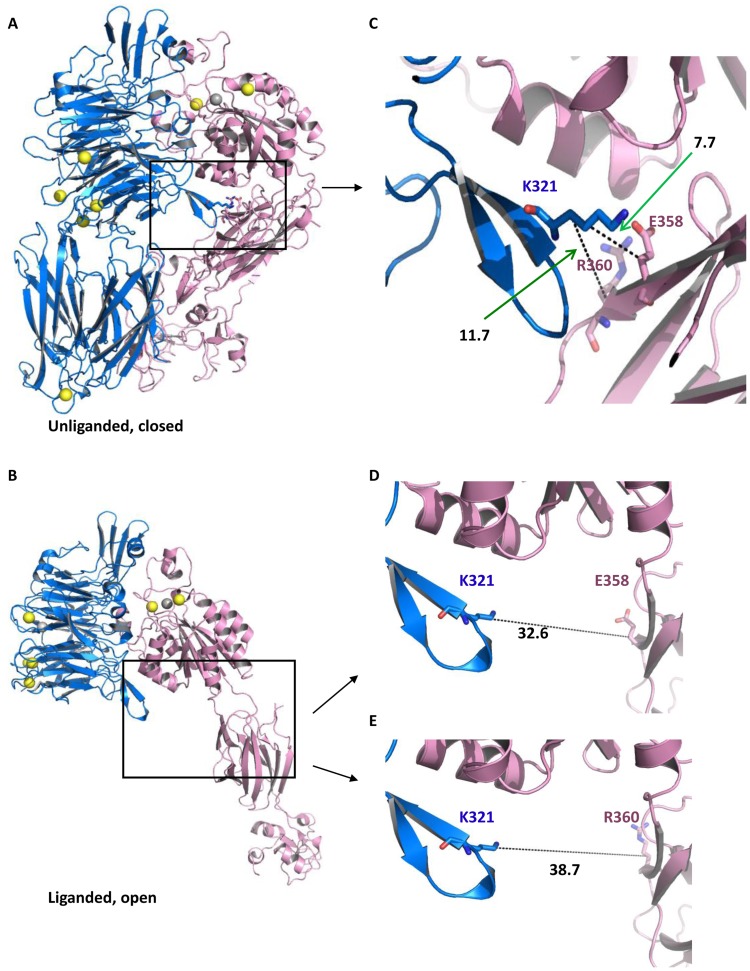
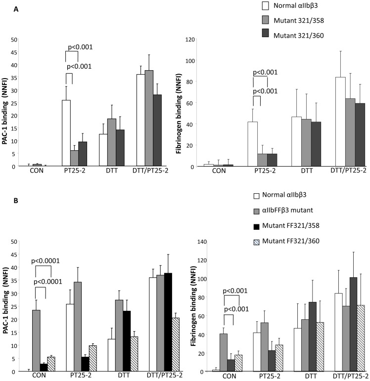
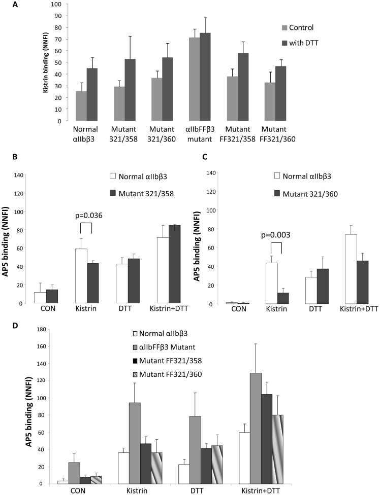
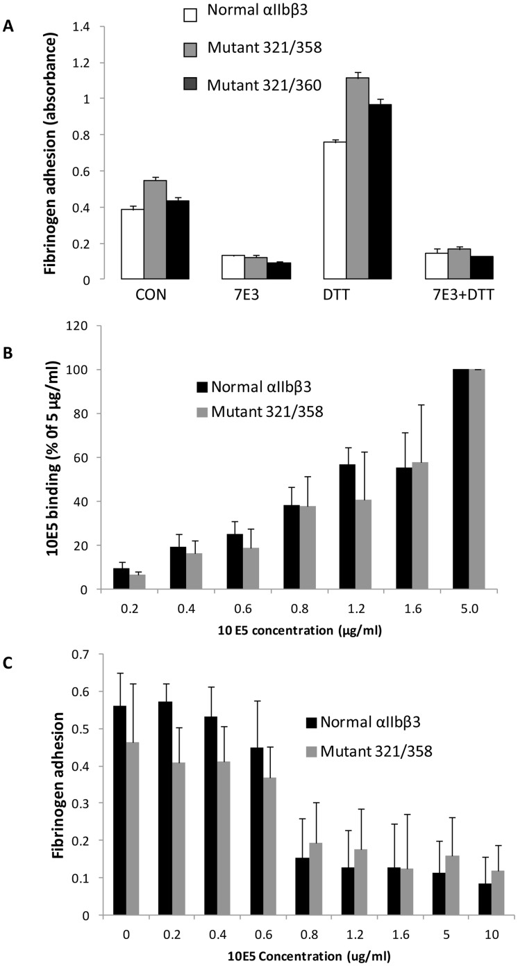
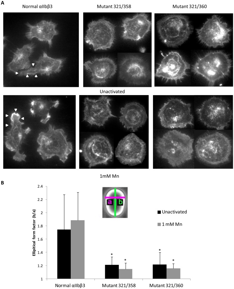
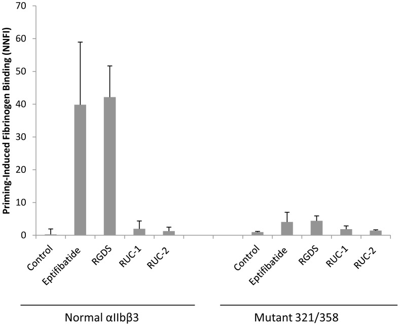
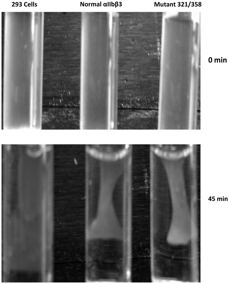
Structural and functional analyses of integrin αIIbβ3 has implicated swing-out motion of the β3 hybrid domain in αIIbβ3 activation and ligand binding. Using data from targeted molecular dynamics (TMD) simulations, we engineered two disulfide-bonded mutant receptors designed to limit swing-out (XS-O). XS-O mutants cannot bind the high Mr ligand fibrinogen in the presence of an activating mAb or after introducing mutations into the αIIb subunit designed to simulate inside-out signaling. They also have reduced capacity to be "primed" to bind fibrinogen by pretreatment with eptifibatide. They can, however, bind the small RGD venom protein kistrin. Despite their inability to bind soluble fibrinogen, the XS-O mutants can support adhesion to immobilized fibrinogen, although such adhesion does not initiate outside-in signaling leading to normal cytoskeletal reorganization. Collectively, our data further define the biologic role of β3 hybrid domain swing-out in both soluble and immobilized high Mr ligand binding, as well as in priming and outside-in signaling. We also infer that swing-out is likely to be a downstream effect of receptor extension.


Conflict of interest statement

Competing Interests: In accordance with Federal Law and the policies of the Research Foundation of the State University of New York, Dr. Barry Coller has a royalty interest in abciximab (Centocor); this does not alter the authors' adherence to all the PLOS ONE policies on sharing data and materials.

Figures

Figure 1
Figure 1. Structures of integrin αIIbβ3 in unliganded, closed and liganded, open conformations.
αIIb subunit is in blue color, β3 subunit is in pink color. A. Unliganded, closed structure of αIIbβ3 (PDB: 3FCS) B. Liganded, open structure of αIIbβ3 (PDB: 3FCU). C. Structural details of unliganded, closed αIIbβ3. The distances between the Cβ atoms of αIIbK321 and β3E358 and between αIIbK321 and β3R360 are 7.7 and 11.7 Å, respectively. D and E. Structural details of the liganded, open αIIbβ3. The distances between the Cβ atoms of αIIbK321and β3E358 and between αIIbK321and β3R360 are 32.6 and 38.7 Å, respectively.
Figure 2
Figure 2. Analysis of mutant protein expression. A. Surface expression of αIIbβ3 on HEK293 cells expressing normal or mutant αIIbβ3 receptors as judged by the binding of mAb 10E5.
1 million cells in 100 µl were incubated with 5 µg/ml Alexa488-conjugated mAb 10E5 (black line) or as a control for non-specific binding, 5 µg/ml Alexa488-conjugated mAb 10E5 in the presence of excess (125 µg/ml) unlabelled 10E5 (gray line; background). B. Disulfide-bonded αIIbβ3 heterodimer formation on the cell surface of XS-O mutants. HEK293 cells expressing normal αIIbβ3 or XS-O mutants were biotinylated and lysed, and lysates were immunoprecipitated with anti-αIIbβ3 complex mAb 10E5. After SDS-PAGE and protein transfer, biotinylated proteins were identified with streptavidin-HRP. Left panel. Non-reduced SDS-PAGE analysis of normal αIIbβ3 and XS-O mutants. Right panel. SDS-PAGE analysis of normal αIIbβ3 and XS-O mutants under reducing conditions (10% β-mercaptoethanol). C and D. Mass spectroscopy identifies unique, predicted peptides from XS-O mutants. C. Purified protein from mutant 321/358 was digested with trypsin and analyzed by LC-MS/MS. A peak at m/z = 488.2569 corresponded to the predicted disulfide linked MH4 3+ ion of peptide VELC(-VR)-CLAEVGR. D. Purified protein from mutant 321/360 was digested with trypsin and ASP-N, followed by LC-MS/MS analysis. A peak at m/z = 653.2639 corresponded to the predicted disulfide linked MH4 + ion of EVC-CLA.
Figure 3
Figure 3. Cells expressing XS-O mutants have reduced ability to bind PAC-1 and fibrinogen.
A. Left, PAC-1 binding of cells expressing XS-O mutants 321/358 and 321/360 the absence of treatment (control; CON) or in the presence of mAb PT25-2, DTT, or DTT+PT25-2. FITC-PAC-1 was added at 5 µg/ml and binding was assessed via flow cytometry. Binding is expressed as net normalized fluorescence intensity (NNFI), in which the geometric mean fluorescence intensity after subtracting nonspecific binding is divided by the relative surface receptor expression as judged by the binding of mAb 10E5. Data expressed as mean ± SD; n = 5. Right, Fibrinogen binding using Alexa488-fibringen (200 µg/ml) as indicated above in “A” for PAC-1 (mean ± SD; n = 6). B. The αIIb F992A/F993A activating mutations fail to rescue PAC-1 and fibrinogen binding in the XS-O mutants.
Figure 4
Figure 4. Kistrin-induced AP5 binding to cells expressing XS-O mutants. A. Kistrin binds to cells expressing XS-O mutants.
Cells expressing normal αIIbβ3 and XS-O mutants were either untreated or treated with DTT (5 mM) before being incubated with Alexa488-kistrin (50 nM). Binding was assessed via flow cytometry and expressed as net normalized fluorescence intensity (mean ± SD; n = 4). B. AP5 binding to mutant 321/358. Alexa488-labeled anti-LIBS mAb AP5 was incubated with cells in the absence or presence of kistrin (200 nM), or DTT (5 mM), or both at 37°C for 1 h. AP5 binding was assessed by flow cytometry and expressed as NNFI, using excess unlabeled AP5 to assess nonspecific binding. C. AP5 binding to XS-O mutant 321/360. D. AP5 binding to αIIbFFβ3 mutant and mutants FF321/358 and FF321/360 (mean ± SD; n = 4).
Figure 5
Figure 5. Cells expressing mutant 321/358 and 321/360 can adhere to immobilized fibrinogen.
A. Cells expressing normal αIIbβ3 or XS-O mutants 321/358 and 321/360 were allowed to adhere to microtiter wells coated with 5 µg/ml fibrinogen without treatment or in the presence of mAb 7E3, DTT, or 7E3+DTT. Adhesion was measured by lysing the washed, adherent cells and assaying for acid phosphatase activity (mean ± SD, n = 4). Relative surface expression of αIIbβ3 based on mAb 10E5 binding in these experiments was 80% for normal αIIbβ3, 100% for 321/358, and 90% for 321/360. B. Binding of mAb 10E5 to cells expressing normal αIIbβ3 or XS-O mutant 321/358. Data are expressed as percentage of the value at 5 µg/ml 10E5. The GMFI values for 10E5 binding at 5 µg/ml were 75±14 for the cells expressing normal αIIbβ3 and 129±39 for the cells expressing the 321/358 mutant (n = 4). C. Adhesion to fibrinogen of cells expressing normal αIIbβ3 or XS-O mutants 321/358 in the presence of different concentrations of mAb 10E5. The same cells used in B. were tested for adhesion to fibrinogen (n = 4).
Figure 6
Figure 6. Cells expressing XS-O mutants 321/358 and 321/360 have defective cytoskeletal reorganization on immobilized fibrinogen.
A. Cells expressing normal αIIbβ3 or XS-O mutants 321/358 or 321/360 were allowed to adhere to microtiter wells coated with 20 µg/ml fibrinogen for 2 hr in HBMT buffer with 2 mM Ca2+ and 1 mM Mg2+. Cells were fixed, permeabilized, and double-stained for actin filaments with rhodamine-phalloidin (red) and β3 with anti-β3 antibody Alex 488-7H2 (green). Image J was used to merge the two color images. Upper panel. Untreated cells. Lower panel. Cells treated with 1 mM Mn2+. B. The eccentricity of cell shape was measured by fitting an ellipse into the image of the cell and measuring both the major and minor axes. Eccentricity was defined as the ratio of the major axis (b) to the minor axis (a) and expressed as the elliptical form factor. * p<0.0001 compared to normal αIIbβ3 (n = 20).
Figure 7
Figure 7. Fibrinogen binding of mutant 321/358 after priming.
HEK293 cells expressing normal αIIbβ3 or mutant 321/358 were incubated with buffer (Control), eptifibatide (1 µM), RGDS (100 µM), RUC-1 (100 µM), RUC-2, (1 µM), or EDTA (10 mM) for 20 min at RT and then fixed and washed 4 times. Binding of Alexa488-conjugated fibrinogen (200 µg/ml) was then assessed by flow cytometry and expressed as NNFI, in which the net geometric mean fluorescence intensity after subtracting the background is divided by the relative surface receptor expression as judged by the binding of mAb 10E5. Data expressed as mean ± SD after subtracting the EDTA value as background (n = 3).
Figure 8
Figure 8. Cells expressing XS-O mutant 321/358 can retract fibrin clots.
Untransduced cells (control 293 cells), cells expressing normal αIIbβ, and cells expressing XS-O mutant 321/358 were incubated with 200 µg/ml fibrinogen and 2 U/ml thrombin at 37°C. Clot retraction was monitored by photography at timed intervals up to 4 hr. The 45 min time point is shown since maximal clot retraction was achieved at this time.

Similar articles

Cited by

References

    1. Campbell ID, Humphries MJ (2011) Integrin structure, activation, and interactions. Cold Spring Harb Perspect Biol 3. - PMC - PubMed
    1. Luo BH, Carman CV, Springer TA (2007) Structural basis of integrin regulation and signaling. Annu Rev Immunol 25: 619–647. - PMC - PubMed
    1. Coller BS, Shattil SJ (2008) The GPIIb/IIIa (integrin alphaIIbbeta3) odyssey: a technology-driven saga of a receptor with twists, turns, and even a bend. Blood 112: 3011–3025. - PMC - PubMed
    1. Coller BS (2001) Anti-GPIIb/IIIa drugs: current strategies and future directions. Thromb Haemost 86: 427–443. - PubMed
    1. Phillips DR, Charo IF, Scarborough RM (1991) GPIIb-IIIa: the responsive integrin. Cell 65: 359–362. - PubMed

MeSH terms

LinkOut - more resources