. 2013 Dec 11;8(12):e81641.
doi: 10.1371/journal.pone.0081641. eCollection 2013.

The simplest integrated multicellular organism unveiled

Affiliations

The simplest integrated multicellular organism unveiled

Yoko Arakaki et al. PLoS One. .

Abstract

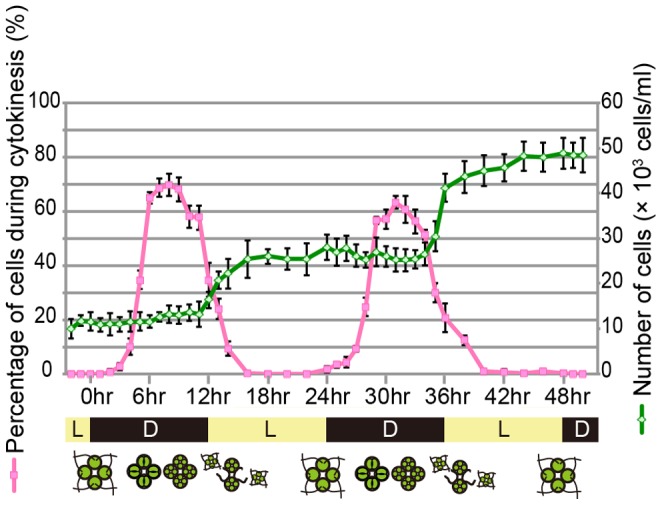
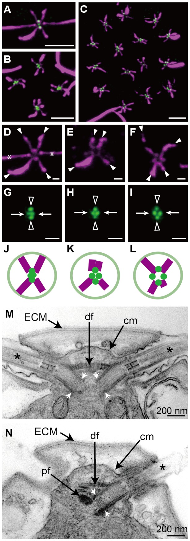
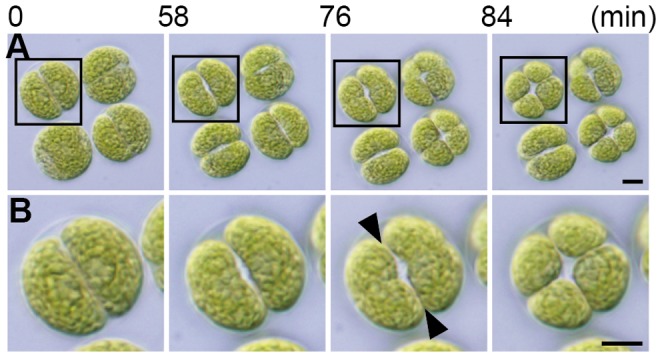
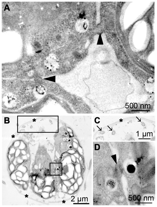
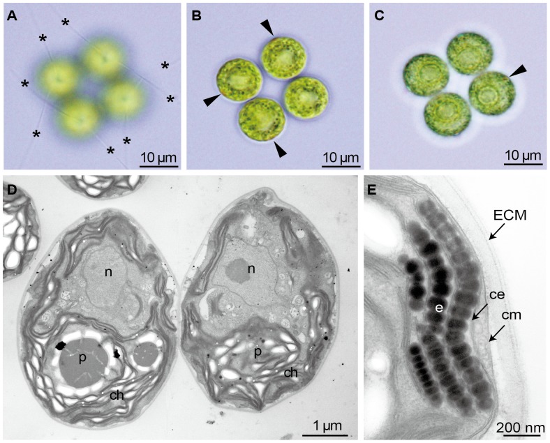
Volvocine green algae represent the "evolutionary time machine" model lineage for studying multicellularity, because they encompass the whole range of evolutionary transition of multicellularity from unicellular Chlamydomonas to >500-celled Volvox. Multicellular volvocalean species including Gonium pectorale and Volvox carteri generally have several common morphological features to survive as integrated multicellular organisms such as "rotational asymmetry of cells" so that the cells become components of the individual and "cytoplasmic bridges between protoplasts in developing embryos" to maintain the species-specific form of the multicellular individual before secretion of new extracellular matrix (ECM). However, these morphological features have not been studied in the four-celled colonial volvocine species Tetrabaena socialis that is positioned in the most basal lineage within the colonial or multicellular volvocine greens. Here we established synchronous cultures of T. socialis and carried out immunofluorescence microscopic and ultrastructural observations to elucidate these two morphological attributes. Based on immunofluorescence microscopy, four cells of the mature T. socialis colony were identical in morphology but had rotational asymmetry in arrangement of microtubular rootlets and separation of basal bodies like G. pectorale and V. carteri. Ultrastructural observations clearly confirmed the presence of cytoplasmic bridges between protoplasts in developing embryos of T. socialis even after the formation of new flagella in each daughter protoplast within the parental ECM. Therefore, these two morphological attributes might have evolved in the common four-celled ancestor of the colonial volvocine algae and contributed to the further increase in cell number and complexity of the multicellular individuals of this model lineage. T. socialis is one of the simplest integrated multicellular organisms in which four identical cells constitute the individual.


Conflict of interest statement

Competing Interests: The authors have declared that no competing interests exist.

Figures

Figure 1
Figure 1. Rough outline of phylogenetic relationships in volvocine green algae , , .
Figure 2
Figure 2. Time course of synchronous culture of Tetrabaena socialis NIES-571.
Light-dark cycle (light:dark = 12 h:12 h) were indicated on the horizontal axis, percentages of cells during cytokinesis were indicated on a vertical line of left side with a pink line, and number of cells were indicated on a right side with a green line. Each error bars shows standard deviation (n = 3).
Figure 3
Figure 3. Images and diagrams of microtubular rootlet (MTR) and basal bodies (BB)/pro-basal bodies (pBB).
(A–I) Immunofluorescence microscopy. (A–C) Double stained fluorescence of acetylated tubulin and CrSAS-6 showing MTR and BB/pBB, respectively. Each scale bar represents 5 µm. (A) Chlamydomonas reinhardtii. (B) Tetrabaena socialis. (C) Gonium pectorale. (D–F) Fluorescence of acetylated tubulin. Each white arrowhead or asterisk indicates distal end of MTR or flagellum, respectively. Each scale bar represents 1 µm. Upper sides of panels E and F represent the directions of center in the flattened colonies. (D) C. reinhardtii. (E) T. socialis. (F) G. pectorale. (G–I) Fluorescence of CrSAS-6. Each arrow or arrowhead indicates BB or pBB, respectively. Each scale bar represents 1 µm. Upper sides of panels H and I represent the directions of center in the flattened colonies. (G) C. reinhardtii. (H) T. socialis. (I) G. pectorale. (J–L) Diagrams of MTR and BB/pBB arrangements. Upper sides of panels K and L represent the directions of center in the flattened colonies. (J) C. reinhardtii. (K) T. socialis. (L) G. pectorale. (M, N) Transmission electron microscopy of T. socialis. ECM, extracellular matrix; cm, cell membrane; df, distal fiber; pf, proximal fiber; asterisk, flagellar proper. (M) Longitudinal section of anterior end of cell showing BB and distal fiber. Note proximal ends of the two BB (white arrows) are separated from each other. (N) Longitudinal section of anterior end of cell showing BB with proximal fiber.
Figure 4
Figure 4. Time-lapse analysis for cytokinesis of Tetrabaena socialis NIES-571.
(A) Parental colony of T. socialis, shown at the same magnification throughout. Scale bar represents 5 µm. (B) Enlarged image in frame in (A), shown at the same magnification throughout. Scale bar represents 5 µm. Note possible connections between daughter protoplasts (arrowheads).
Figure 5
Figure 5. Transmission electron microscopy of cytokinesis of four-celled embryos of Tetrabaena socialis NIES-571.
(A) Transverse section of almost central part of four daughter protoplasts before formation of new flagella. Note a daughter protoplast connected to two neighbors by cytoplasmic bridges (arrowheads). (B) Semi-longitudinal section of daughter protoplasts after formation of new flagella (large frame) within parental extracellular matrix (ECM) (asterisks). Note two protoplast connected to each other by cytoplasmic bridges (small frame). (C, D) Enlarged images of two frames in (B). (C) New flagella (arrows) within parental ECM (asterisks). (D) Cytoplasmic bridges (arrowhead) connecting two daughter protoplasts.
Figure 6
Figure 6. Light and transmission electron microscopy of vegetative colonies of Tetrabaena socialis NIES-571.
(A–C) Three light microscopic views of four-celled vegetative colony, showing positions of flagella and eyespots. Note that flagella (asterisks) and eyespots (arrowheads) are arranged in symmetric pattern in the whole colony. (D, E) Transmission electron microscopy. ECM, extracellular matrix; ce, chloroplast envelope; ch, chloroplast; cm, cell membrane; n, nucleus; p, pyrenoid. (D) Longitudinal section of vegetative colony. (E) Eyespot composed three layers of globules.

References

    1. Michod RE (2005) On the transfer of fitness from the cell to the multicellular organism. Biol Philos 20: 967–987.
    1. Abedin M, King N (2010) Diverse evolutionary paths to cell adhesion. Trends Cell Biol 20: 734–742. - PMC - PubMed
    1. Grosberg RK, Strathmann RR (2007) The evolution of multicellularity: a minor major transition? Annu Rev Ecol Evol 38: 621–654.
    1. Kirk DL (2005) A twelve-step program for evolving multicellularity and a division of labor. BioEssays 27: 299–310. - PubMed
    1. Sachs JL (2008) Resolving the first steps to multicellularity. Trends Ecol Evol 23: 245–248. - PubMed

Publication types

LinkOut - more resources