New therapeutic approach: diphenyl diselenide reduces mitochondrial dysfunction in acetaminophen-induced acute liver failure
- PMID: 24349162
- PMCID: PMC3859582
- DOI: 10.1371/journal.pone.0081961
New therapeutic approach: diphenyl diselenide reduces mitochondrial dysfunction in acetaminophen-induced acute liver failure
Abstract
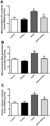
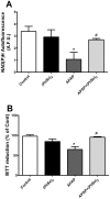
The acute liver failure (ALF) induced by acetaminophen (APAP) is closely related to oxidative damage and depletion of hepatic glutathione, consequently changes in cell energy metabolism and mitochondrial dysfunction have been observed after APAP overdose. Diphenyl diselenide [(PhSe)2], a simple organoselenium compound with antioxidant properties, previously demonstrated to confer hepatoprotection. However, little is known about the protective mechanism on mitochondria. The main objective of this study was to investigate the effects (PhSe)2 to reduce mitochondrial dysfunction and, secondly, compare in the liver homogenate the hepatoprotective effects of the (PhSe)2 to the N-acetylcysteine (NAC) during APAP-induced ALF to validate our model. Mice were injected intraperitoneal with APAP (600 mg/kg), (PhSe)2 (15.6 mg/kg), NAC (1200 mg/kg), APAP+(PhSe)2 or APAP+NAC, where the (PhSe)2 or NAC treatment were given 1 h following APAP. The liver was collected 4 h after overdose. The plasma alanine and aspartate aminotransferase activities increased after APAP administration. APAP caused a remarkable increase of oxidative stress markers (lipid peroxidation, reactive species and protein carbonylation) and decrease of the antioxidant defense in the liver homogenate and mitochondria. APAP caused a marked loss in the mitochondrial membrane potential, the mitochondrial ATPase activity, and the rate of mitochondrial oxygen consumption and increased the mitochondrial swelling. All these effects were significantly prevented by (PhSe)2. The effectiveness of (PhSe)2 was similar at a lower dose than NAC. In summary, (PhSe)2 provided a significant improvement to the mitochondrial redox homeostasis and the mitochondrial bioenergetics dysfunction caused by membrane permeability transition in the hepatotoxicity APAP-induced.
Conflict of interest statement
Figures


Similar articles
-
Acute brain damage induced by acetaminophen in mice: effect of diphenyl diselenide on oxidative stress and mitochondrial dysfunction.Neurotox Res. 2012 Apr;21(3):334-44. doi: 10.1007/s12640-011-9288-1. Epub 2011 Nov 12. Neurotox Res. 2012. PMID: 22081409
-
Reversal of bioenergetics dysfunction by diphenyl diselenide is critical to protection against the acetaminophen-induced acute liver failure.Life Sci. 2017 Jul 1;180:42-50. doi: 10.1016/j.lfs.2017.05.012. Epub 2017 May 10. Life Sci. 2017. PMID: 28501483
-
Reduction of acute hepatic damage induced by acetaminophen after treatment with diphenyl diselenide in mice.Toxicol Pathol. 2012 Jun;40(4):605-13. doi: 10.1177/0192623311436179. Epub 2012 Feb 1. Toxicol Pathol. 2012. PMID: 22301948
-
Mechanisms of acetaminophen-induced liver injury and its implications for therapeutic interventions.Redox Biol. 2018 Jul;17:274-283. doi: 10.1016/j.redox.2018.04.019. Epub 2018 Apr 22. Redox Biol. 2018. PMID: 29753208 Free PMC article. Review.
-
The molecular mechanisms of acetaminophen-induced hepatotoxicity and its potential therapeutic targets.Exp Biol Med (Maywood). 2023 May;248(5):412-424. doi: 10.1177/15353702221147563. Epub 2023 Jan 20. Exp Biol Med (Maywood). 2023. PMID: 36670547 Free PMC article. Review.
Cited by
-
Acute Exposure to Permethrin Modulates Behavioral Functions, Redox, and Bioenergetics Parameters and Induces DNA Damage and Cell Death in Larval Zebrafish.Oxid Med Cell Longev. 2019 Nov 11;2019:9149203. doi: 10.1155/2019/9149203. eCollection 2019. Oxid Med Cell Longev. 2019. PMID: 31827707 Free PMC article.
-
Label-Free Imaging Techniques to Evaluate Metabolic Changes Caused by Toxic Liver Injury in PCLS.Int J Mol Sci. 2023 May 24;24(11):9195. doi: 10.3390/ijms24119195. Int J Mol Sci. 2023. PMID: 37298155 Free PMC article.
-
Functional roles of protein nitration in acute and chronic liver diseases.Oxid Med Cell Longev. 2014;2014:149627. doi: 10.1155/2014/149627. Epub 2014 Apr 30. Oxid Med Cell Longev. 2014. PMID: 24876909 Free PMC article. Review.
-
4-Hydroxyphenylacetic Acid Prevents Acute APAP-Induced Liver Injury by Increasing Phase II and Antioxidant Enzymes in Mice.Front Pharmacol. 2018 Jun 19;9:653. doi: 10.3389/fphar.2018.00653. eCollection 2018. Front Pharmacol. 2018. PMID: 29973881 Free PMC article.
-
N-Acetyl Cysteine Overdose Inducing Hepatic Steatosis and Systemic Inflammation in Both Propacetamol-Induced Hepatotoxic and Normal Mice.Antioxidants (Basel). 2021 Mar 12;10(3):442. doi: 10.3390/antiox10030442. Antioxidants (Basel). 2021. PMID: 33809388 Free PMC article.
References
-
- Larson AM, Polson J, Fontana RJ, Davern TJ, Lalani E, et al. (2005) Acetaminophen-induced acute liver failure: results of a United States multicenter, prospective study. Hepatology 42: 1364–1372. - PubMed
-
- Nourjah P, Ahmad SR, Karwoski C, Willy M (2006) Estimates of acetaminophen (paracetamol)-associated overdoses in the United States. Pharmacoepidemiology and Drug Safety 15: 398–405. - PubMed
-
- de Achaval S, Suarez-Almazor M (2011) Acetaminophen overdose: a little recognized public health threat. Pharmacoepidemiology and Drug Safety 20: 827–829. - PubMed
-
- Brown JM, Ball JG, Hogsett A, Williams T, Valentovic M (2010) Temporal study of acetaminophen (APAP) and S-adenosyl-L-methionine (SAMe) effects on subcellular hepatic SAMe levels and methionine adenosyltransferase (MAT) expression and activity. Toxicology and Applied Pharmacology 247: 1–9. - PMC - PubMed
MeSH terms
Substances
LinkOut - more resources
Full Text Sources
Other Literature Sources
Medical

