. 2013 Dec 11;8(12):e81982.
doi: 10.1371/journal.pone.0081982. eCollection 2013.

Carnosine's effect on amyloid fibril formation and induced cytotoxicity of lysozyme

Affiliations

Carnosine's effect on amyloid fibril formation and induced cytotoxicity of lysozyme

Josephine W Wu et al. PLoS One. .

Abstract

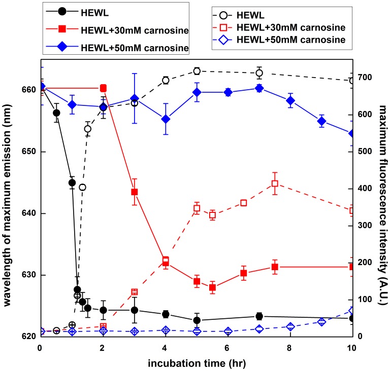
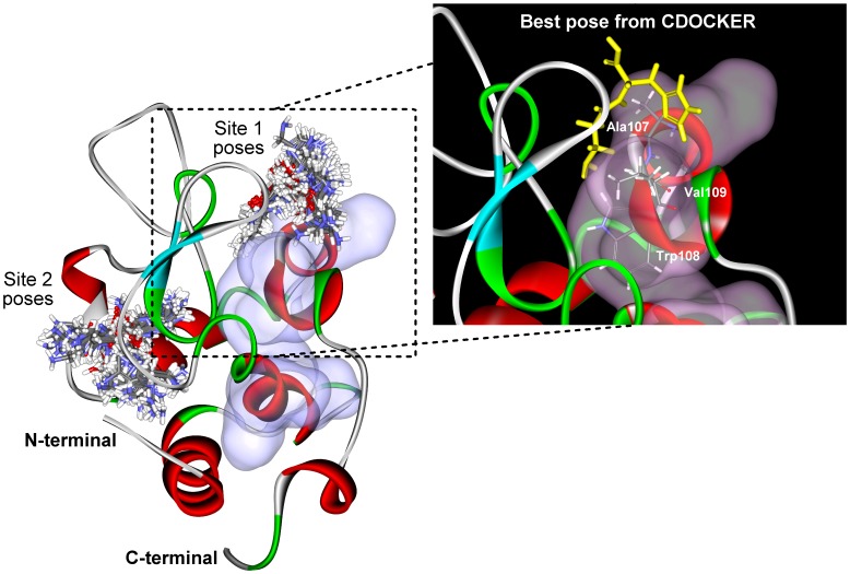
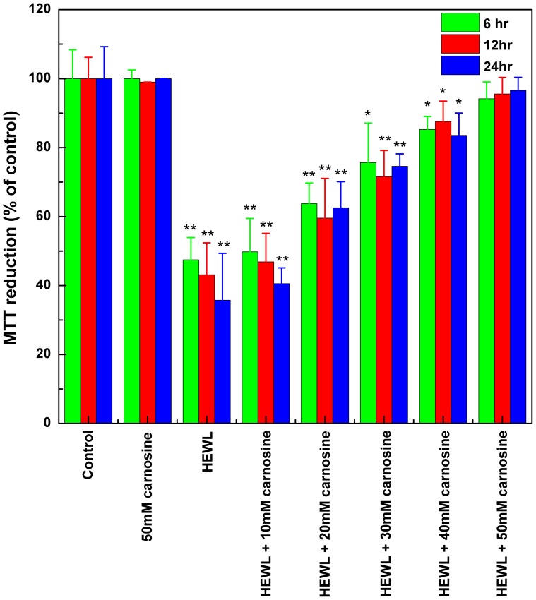
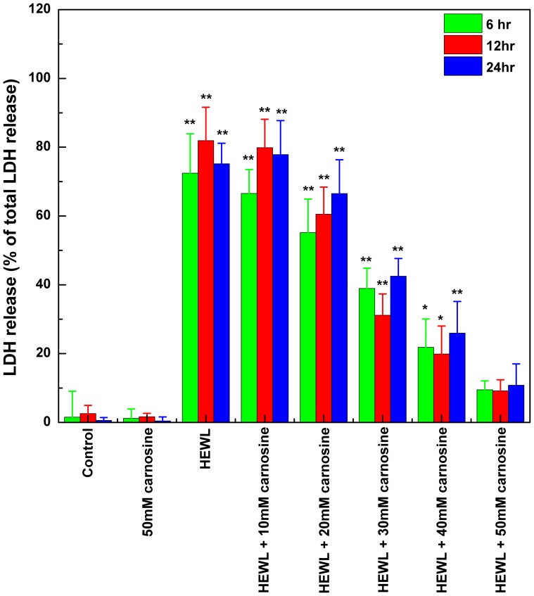
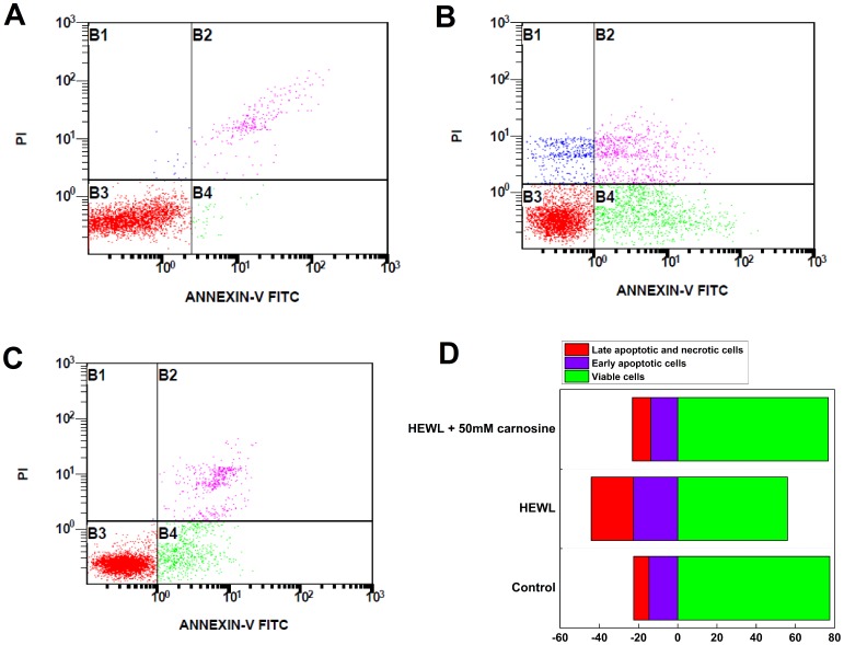
Carnosine, a common dipeptide in mammals, has previously been shown to dissemble alpha-crystallin amyloid fibrils. To date, the dipeptide's anti-fibrillogensis effect has not been thoroughly characterized in other proteins. For a more complete understanding of carnosine's mechanism of action in amyloid fibril inhibition, we have investigated the effect of the dipeptide on lysozyme fibril formation and induced cytotoxicity in human neuroblastoma SH-SY5Y cells. Our study demonstrates a positive correlation between the concentration and inhibitory effect of carnosine against lysozyme fibril formation. Molecular docking results show carnosine's mechanism of fibrillogenesis inhibition may be initiated by binding with the aggregation-prone region of the protein. The dipeptide attenuates the amyloid fibril-induced cytotoxicity of human neuronal cells by reducing both apoptotic and necrotic cell deaths. Our study provides solid support for carnosine's amyloid fibril inhibitory property and its effect against fibril-induced cytotoxicity in SH-SY5Y cells. The additional insights gained herein may pave way to the discovery of other small molecules that may exert similar effects against amyloid fibril formation and its associated neurodegenerative diseases.


Conflict of interest statement

Competing Interests: The authors have declared that no competing interests exist.

Figures

Figure 1
Figure 1. Amyloid fibrillogenesis kinetics of hen egg-white lysozyme (HEWL).
The extent of fibril formation was monitored via Congo red absorbance at 540(ThT) fluorescence as a function of incubation time. HEWL samples were dissolved in 100 mM glycine buffer (pH 2.0) and incubated at 55°C with agitation of 580 rpm during the course of experiment. The extent of fibril formation was also monitored via Congo red absorption spectra shown in the inset (the arrow indicates the increasing incubation time). Data represent the mean ThT fluorescence intensity of at least 5 independent experiments (n≥5). Error bars represent the standard deviation (S.D.) of the fluorescence measurements.
Figure 2
Figure 2. Effects of carnosine on the kinetics of amyloid fibrillogenesis of HEWL.
Inhibition of HEWL fibril formation by carnosine in a concentration-dependent manner as revealed by (A) ThT fluorescence and (B and C) Congo red absorbance. The Congo red binding absorbance spectra of HEWL samples were taken at (B) 0 hr and (C) 10 hr of incubation. The samples with and without carnosine were dissolved in 100 mM glycine buffer (pH 2.0) and incubated at 55°C agitated at 580 rpm during the course of the experiment. Each data point represents the mean value of at least 5 independent experiments (n≥5). Error bars denote the standard deviation (S.D.) of the measurements.
Figure 3
Figure 3. Transmission electron micrographs of HEWL samples with various concentrations of carnosine.
Negatively stained electron micrographs of (A) HEWL alone, (B) HEWL co-incubated with 20 mM carnosine, (C) HEWL co-incubated with 40 mM carnosine, and (D) HEWL co-incubated with 50 mM carnosine. HEWL samples in the absence and presence of carnosine were prepared at pH 2.0 and incubated for 10 hr. The scale bar represents 50 nm.
Figure 4
Figure 4. Representative far-UV CD spectra of HEWL samples.
The far-UV CD spectra of (A) HEWL alone, (B) HEWL co-incubated with 10 mM carnosine, (C) HEWL co-incubated with 30 mM carnosine, and (D) HEWL co-incubated with 50 mM carnosine. The inset illustrates changes in HEWL secondary structure, showing the contents of α-helix and β-sheet during the fibril formation process.
Figure 5
Figure 5. The effects of carnosine on the hydrophobicity of HEWL during incubation.
The time-course of the protein surface hydrophobicity was measured by Nile red-binding fluorescence at different incubation times. Data were presented as maximum Nile red fluorescence intensity and wavelength of maximum fluorescence emission taken at various incubation time. Each point represents the average of at least 5 independent measurements (n≥5). HEWL samples were dissolved in 100 mM glycine buffer (pH 2.0) and incubated at 55°C accompanied by agitation of 580 rpm during the course of the experiment (solid symbols represent the wavelength of maximum fluorescence and open symbols are indicative of maximum Nile red fluorescence intensity).
Figure 6
Figure 6. Docking poses of carnosine generated for each of the two predicted binding sites (Sites 1 and 2).
The 20-prone regions predicted by the bioinformatics aggregation predictors are surface rendered in violet. Inset: the best docked pose (Pose 1; 0.2 Å yellow-colored sticks) at Site 1 and the three interacting residues (0.12 Å atom-colored sticks) of aggregation-prone region.
Figure 7
Figure 7. Carnosine's effect on SH-SY5Y cell viability.
The cell viability upon exposure to various HEWL samples was measured by MTT reduction. The SH-SY5Y cells were exposed to 50 mM carnosine alone (the negative control) and HEWL samples aged for 10 hr without or with various concentrations of carnosine (10, 20, 30, 40, and 50 mM) for 6, 12, and 24 hr at 37°C in a humidified 5% (v/v) CO2/air environment. The data are presented as the percentage of MTT reduced by the cells incubated in media containing 50 mM carnosine alone, HEWL alone, or HEWL with various concentrations of carnosine. The means ± S.D. of at least 10 determinations are shown.
Figure 8
Figure 8. The effects of carnosine on HEWL-induced membrane damage (LDH release into the medium) in SH-SY5Y cells.
The cell viability upon exposure to HEWL sample was measured by the LDH release assay. SH-SY5Y cells were incubated with 50 mM carnosine alone (the negative control) and HEWL samples without or with various concentrations of carnosine (10, 20, 30, 40, and 50 mM) for 6, 12, and 24 hr at 37°C in a humidified 5% (v/v) CO2/air environment. The percentage of cytotoxicity was evaluated as a ratio of the quantity of LDH released in each sample divided by the total LDH released by the sample of cells treated with lysis buffer. Quantity of released LDH is estimated by the activity of lactate dehydrogenase in the suspension aliquot from the 96-well plates after 30 min incubation with the appropriate substrate solution. Measurements of the means ± S.D. of at least 8 determinations for each sample were obtained at 490 nm.
Figure 9
Figure 9. The effects of carnosine on aged HEWL-induced cell deaths analyzed by flow cytometry.
Representative intensity dot plots of (A) un-treated SH-SY5Y cells (the un-treated control group); (B) SH-SY5Y cells co-incubated with 10-hr aged HEWL sample; (C) SH-SY5Y cells co-incubated with 10-hr aged HEWL sample containing 50 mM carnosine. The dotted plots are divided into viable (bottom left quadrants; Annexin V-FITC/PI double negative), early apoptotic (bottom right quadrants; Annexin V-FITC positive, PI negative), and late apoptotic/necrotic cells (top right quadrants; Annexin V-FITC/PI double positive) according to Annexin V-FITC and PI fluorescence. (D) Summary of the effects of carnosine on aged HEWL-induced apoptosis. Results are presented as the percentages of viable cells (Annexin V-FITC/PI double negative), early apoptotic cells (Annexin V-FITC positive, PI negative), and late apoptotic/necrotic cells (Annexin V-FITC FITC/PI double positive) according to the PI fluorescence versus Annexin V-FITC fluorescence dotted plots shown in (A), (B), and (C).

References

    1. Dobson CM (2004) Principles of protein folding, misfolding and aggregation. Semin Cell Dev Biol 15: 3–16. - PubMed
    1. Wang SSS, Good TA (2005) An overview of Alzheimer's disease. Journal of the Chinese Institute of Chemical Engineers 36: 533–559.
    1. Liberski PP, Sikorska B, Hauw JJ, Kopp N, Streichenberger N, et al. (2010) Ultrastructural Characteristics (or Evaluation) of Creutzfeldt-Jakob Disease and Other Human Transmissible Spongiform Encephalopathies or Prion Diseases. Ultrastructural Pathology 34: 351–361. - PubMed
    1. Kell DB (2010) Towards a unifying, systems biology understanding of large-scale cellular death and destruction caused by poorly liganded iron: Parkinson's, Huntington's, Alzheimer's, prions, bactericides, chemical toxicology and others as examples. Archives of Toxicology 84: 825–889. - PMC - PubMed
    1. Ross CA, Poirier MA (2004) Protein aggregation and neurodegenerative disease. Nat Med 10 SupplS10–17. - PubMed

MeSH terms

LinkOut - more resources