Clinical Trial
. 2013 Dec 12;8(12):e82078.
doi: 10.1371/journal.pone.0082078. eCollection 2013.

Electrical vestibular stimulation after vestibular deafferentation and in vestibular schwannoma

Affiliations
Clinical Trial

Electrical vestibular stimulation after vestibular deafferentation and in vestibular schwannoma

Swee Tin Aw et al. PLoS One. .

Abstract

Background: Vestibular reflexes, evoked by human electrical (galvanic) vestibular stimulation (EVS), are utilized to assess vestibular function and investigate its pathways. Our study aimed to investigate the electrically-evoked vestibulo-ocular reflex (eVOR) output after bilateral and unilateral vestibular deafferentations to determine the characteristics for interpreting unilateral lesions such as vestibular schwannomas.

Methods: EVOR was recorded with dual-search coils as binocular three-dimensional eye movements evoked by bipolar 100 ms-step at EVS intensities of [0.9, 2.5, 5.0, 7.5, 10.0] mA and unipolar 100 ms-step at 5 mA EVS intensity. Five bilateral vestibular deafferented (BVD), 12 unilateral vestibular deafferented (UVD), four unilateral vestibular schwannoma (UVS) patients and 17 healthy subjects were tested with bipolar EVS, and five UVDs with unipolar EVS.

Results: After BVD, bipolar EVS elicited no eVOR. After UVD, bipolar EVS of one functioning ear elicited bidirectional, excitatory eVOR to cathodal EVS with 9 ms latency and inhibitory eVOR to anodal EVS, opposite in direction, at half the amplitude with 12 ms latency, exhibiting an excitatory-inhibitory asymmetry. The eVOR patterns from UVS were consistent with responses from UVD confirming the vestibular loss on the lesion side. Unexpectedly, unipolar EVS of the UVD ear, instead of absent response, evoked one-third the bipolar eVOR while unipolar EVS of the functioning ear evoked half the bipolar response.

Conclusions: The bidirectional eVOR evoked by bipolar EVS from UVD with an excitatory-inhibitory asymmetry and the 3 ms latency difference between normal and lesion side may be useful for detecting vestibular lesions such as UVS. We suggest that current spread could account for the small eVOR to 5 mA unipolar EVS of the UVD ear.


Conflict of interest statement

Competing Interests: GM Halmagyi and KP Weber are unpaid consultants for GN Otometrics in the video head impulse system. N Lehnen is a shareholder of EyeSeeTec, video head impulse system. No GN Otometrics or EyeSeeTec equipment was used in the study. This does not alter the authors' adherence to all the PLOS ONE policies on sharing data and materials.

Figures

Figure 1
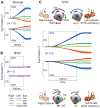
Figure 1. EVORs to human bipolar EVS in healthy subjects, bilateral and unilateral vestibular deafferented patients.
(A) Normal eVOR from healthy subjects (N = 17) (group means ± SEM) comprised conjugate torsional and horizontal eye rotations, binocularly equal in amplitude, rotated away from cathode towards anode, and vertical divergence with the intorting eye upwards (cathode side) and extorting eye downwards (anode side). (B) EVOR was absent from BVD patients (N = 5) with torsional, vertical and horizontal eVOR positions ≤0.01°. (C) Bidirectional eVOR from grouped as right UVD patients with left functioning ear (N = 12). The excitatory eVOR to left-cathode/right-anode (lc/ra) cathodal EVS comprised conjugate torsional and horizontal eye rotations away from cathode and a vertical divergence with the eye on the anodal side moving downwards. The inhibitory eVOR to right-cathode/left-anode (rc/la) anodal EVS was in the opposite direction and at about half the amplitude of the excitatory eVOR with the eye on the anodal side moving upwards. (The schemes illustrate EVS polarities and eye rotation directions).
Figure 2
Figure 2. Spatio-temporal characteristics of the eVOR to human bipolar EVS.
(A, B) Mean eVOR velocity and acceleration time-series to EVS intensities of [0.9, 2.5, 5.0, 7.5, 10.0]mA from grouped as right UVDs with functioning left ears (N = 12). Tonic and phasic eVORs graded to all current intensities for lc/ra cathodal EVS excitation or rc/la anodal EVS inhibition exhibiting excitatory-inhibitory asymmetries. (C) Relationship of eVOR with EVS duration and frequency. Normal eVOR was linearly correlated with EVS duration when EVS duration was incremented from 10–100 ms in 10 ms-step. (D) Comparison of excitatory and inhibitory eVOR positions to a 1 ms EVS step at [1, 50, 100, 200]Hz and to a 100 ms EVS step at 1 Hz. We found that the eVOR correlated well to EVS duration but not to frequency of stimulation.
Figure 3
Figure 3. Latency of the eVOR after UVD.
(A) Excitatory and inhibitory eVOR latencies of binocular torsional, vertical and horizontal eVOR to 5.0 mA lc/ra cathodal EVS and rc/la anodal EVS from grouped as right UVDs with functioning left ears (N = 12, group means ± SEM) showing their mean latencies. (B) Schematic depicting the excitatory (solid red line) and inhibitory (dashed red line) horizontal semicircular canal pathways showing the 3-neuron reflex arc comprising 1: vestibular nerve; 2: vestibulo-ocular secondary neuron; 3: abducens motorneuron. LR: lateral rectus muscle; MR: medial rectus muscle; ON: oculomotor nucleus; AN: abducens nucleus; VN: vestibular nucleus; MLF: medial longitudinal fasciculus. Equivalent 3-neuron reflex arcs also exist for the vertical semicircular canal pathways.
Figure 4
Figure 4. Effect of current spread in human unipolar EVS of UVD patients.
The eVOR to unipolar 5.0(A) The eVORs from left-cathode/right-anode (lc/ra) and to right-cathode/left-anode (rc/la) bipolar EVS were largest. (B) When left-cathode/C7-anode (lc/C7a) and C7-cathode/left-anode (C7c/la) unipolar EVS stimulated the functioning left ear, the eVOR was about half the bipolar eVOR. (C) However when right-cathode/C7-anode (rc/C7a) and C7-cathode/right-anode (C7c/ra) unipolar EVS stimulated the right UVD ear, instead of the expected absent response, it was one-third the bipolar eVOR. The polarity of the eVOR was consistent with the C7- electrode polarity suggesting that current may have spread from the C7-electrode closer to functioning left ear. (D) When the eVOR from all three configurations were grouped according to direction of their responses (i.e. CW or CCW) and then normalized, they share similar spatio-temporal characteristics with the mean excitatory eVOR latency of 8.9 ms and inhibitory latency of 11.4 ms suggesting that the eVORs were generated from the functioning left ear. (Schemes show electrode locations on the patient).
Figure 5
Figure 5. Comparison of the mean eVOR in UVS to normal and UVD subjects.
(A) The mean eVOR in UVS (N = 4) was similar to UVD grouped as right lesions, but different from normal. Right UVS showed bidirectional, excitatory eVOR to 5 mA cathodal EVS and inhibitory eVOR to anodal EVS of the left ear at half the amplitude and opposite in direction, with excitatory-inhibitory asymmetrical response pattern similar to the right UVD. Mean latencies from 4 individual UVS patients (P1, P2, P3, P4) were compared to normal and right UVD subjects. (C) MRI from UVS patients (P1, P2)

References

    1. Purkinje JE. (1823) Commentatio de examine physiologico organi visus et systematis cutanei. Vratislavae, Typis Universitatis.
    1. Politzer A (1909) In: Ballin MJ, Heller CL, editors. Diseases of the ear, fifth edition, Bailliere, Tindall and Cox, London. pp. 90–92.
    1. Goldberg JM, Smith CE, Fernandez C (1984) Relation between discharge regularity and responses to externally applied galvanic currents in vestibular nerve afferents of the squirrel monkey. J Neurophysiol 51: 1236–1256. - PubMed
    1. Aw ST, Todd MJ, Curthoys IS, Aw GE, McGarvie LA, et al... (2010) Vestibulo-ocular responses to sound, vibration and galvanic stimulation. In: Eggers SDZ, Zee DS, editors. Vertigo and Imbalance: Clinical Neurophysiology of the Vestibular System. Handbook of Clinical Neurophysiology. Elsevier Vol 9. pp. 165–180.
    1. Vailleau B, Qu'hen C, Vidal PP, de Waele C (2011) Probing residual vestibular function with galvanic stimulation in vestibular loss patients. Otol Neurotol 32: 863–71. - PubMed

Publication types