. 2013 Dec 12;8(12):e82143.
doi: 10.1371/journal.pone.0082143. eCollection 2013.

Skeletal cell differentiation is enhanced by atmospheric dielectric barrier discharge plasma treatment

Affiliations

Skeletal cell differentiation is enhanced by atmospheric dielectric barrier discharge plasma treatment

Marla J Steinbeck et al. PLoS One. .

Abstract

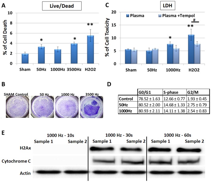
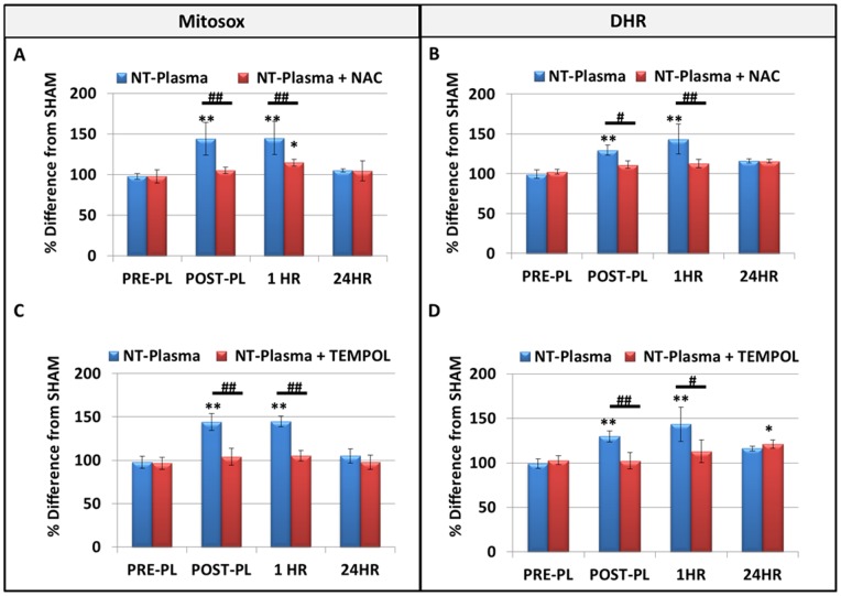
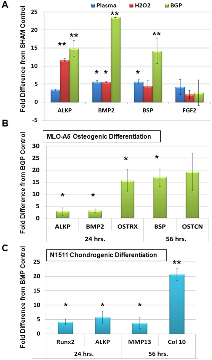
Enhancing chondrogenic and osteogenic differentiation is of paramount importance in providing effective regenerative therapies and improving the rate of fracture healing. This study investigated the potential of non-thermal atmospheric dielectric barrier discharge plasma (NT-plasma) to enhance chondrocyte and osteoblast proliferation and differentiation. Although the exact mechanism by which NT-plasma interacts with cells is undefined, it is known that during treatment the atmosphere is ionized generating extracellular reactive oxygen and nitrogen species (ROS and RNS) and an electric field. Appropriate NT-plasma conditions were determined using lactate-dehydrogenase release, flow cytometric live/dead assay, flow cytometric cell cycle analysis, and Western blots to evaluate DNA damage and mitochondrial integrity. We observed that specific NT-plasma conditions were required to prevent cell death, and that loss of pre-osteoblastic cell viability was dependent on intracellular ROS and RNS production. To further investigate the involvement of intracellular ROS, fluorescent intracellular dyes Mitosox (superoxide) and dihydrorhodamine (peroxide) were used to assess onset and duration after NT-plasma treatment. Both intracellular superoxide and peroxide were found to increase immediately post NT-plasma treatment. These increases were sustained for one hour but returned to control levels by 24 hr. Using the same treatment conditions, osteogenic differentiation by NT-plasma was assessed and compared to peroxide or osteogenic media containing β-glycerolphosphate. Although both NT-plasma and peroxide induced differentiation-specific gene expression, neither was as effective as the osteogenic media. However, treatment of cells with NT-plasma after 24 hr in osteogenic or chondrogenic media significantly enhanced differentiation as compared to differentiation media alone. The results of this study show that NT-plasma can selectively initiate and amplify ROS signaling to enhance differentiation, and suggest this technology could be used to enhance bone fusion and improve healing after skeletal injury.


Conflict of interest statement

Competing Interests: The authors have declared that no competing interests exist.

Figures

Figure 1
Figure 1. Direct effects of NT-plasma on cytotoxicty and cell proliferation.
Osteoblast-like, MLO-A5 cells were treated with NT-plasma at frequencies of 50, 1000 and 3500 Hz for 10 seconds. (A) Cell death measured by nuclear PI incorporation at 24 hr showed increased cell death in response to frequency 50 Hz, 3500 Hz (p<0.01) and H2O2 (p<0.001) (n = 3). There was no significant cell death in response to frequency 1000 Hz. (B) Cell detachment was observed at 3500 Hz as shown by toluidine staining of cells after NT-plasma treatment. (C) Cell viability after NT-plasma was assessed by a lactate dehydrogenase (LDH) release assay. 1000 Hz and H2O2 both showed an increase in LDH release (p<0.01 and p<0.001). LDH release was reduced in the presence of TEMPOL for both H2O2 (p<0.01) and NT-plasma treatments (ns, n = 3). (D) No difference in cell cycle profile was observed between NT-plasma at 50 Hz or 1000 Hz as compared to sham control. (E) Western blots show no H2Ax or cytochrome c release in cells treated at 1000 Hz for 10 s as compared to the 30 and 60 sec NT-plasma treatments. Statistical significance was determined by the Mann-Whitney U test for non-parametric data; * or # (p<0.01) ** or ## (p<0.001.
Figure 2
Figure 2. Direct effects of NT-plasma on intracellular ROS production.
(A and B) Intracellular O2 . and H2O2 levels were measured using fluorescent indicators MitoSox (n = 3) and dihydrorhodamine (DHR), (n = 3) respectively. Immediately post-treatment (POST-PL) and at 1 hr, 1000 Hz NT-plasma treated cells generated significantly increased amounts of O2 . and H2O2 (p<0.001) as compared to sham control or pre-treatment levels (PRE-PL). Amounts of both ROS were decreased by 24 hr. NAC and TEMPOL quenched the ROS increase POST-PL and at 1 hr (p<0.01–0.001). However, H2O2 levels in the TEMPOL (p<0.01) inhibitor group were significantly increased above control at 24 hr, presumably due to removal of baseline NO. The results are expressed as the mean ± standard deviation. Statistical significance was determined by the Mann-Whitney U test for non-parametric data; * or # (p<0.01) ** or ## (p<0.001.
Figure 3
Figure 3. NT-plasma induced osteogenic differentiation using qPCR markers.
(A) Fold increases in the expression of alkaline phosphatase (ALKP), bone morphogenetic protein 2 (BMP2), bone sialoprotein (BSP) and fibroblast growth factor-2 (FGF-2) in response to NT-plasma treatment, H2O2 or β-glycerol phosphate (βGP) normalized to sham treated control cells after 24 hr. ALKP was upregulated 3- (NS) and 11-fold (p<0.01), respectively, and BMP2 and BSP were increased 5 fold (p<0.05). βGP increased expression 14–23 fold (p<0.01). FGF-2 expression was not affected by NT-plasma, H2O2 or βGP. (B) After a 24 hr incubation in βGP NT-plasma was applied. 24 hr later there was a 2–7 fold increase in the induction of BMP2, ALKP (p<0.05) and Osterix was increased 15 fold (p<0.01) compared to βGP treatment alone. At 56 hr, BSP and osteocalcin (OSTCN) were increased 17–24 fold above βGP-treated control (p<0.05). The results are expressed as the mean ± standard deviation (n = 2). Statistical significance was determined by the Wilcoxin Mann-Whitney test for non-parametric data; * (p<0.05), ** (p<0.01).

References

    1. Glover JL, Bendick PJ, Link WJ, Plunkett RJ (1982) The plasma scalpel: a new thermal knife. Lasers Surg Med 2: 101–106. - PubMed
    1. Arjunan KP, Friedman G, Fridman A, Clyne AM (2011) Non-thermal dielectric barrier discharge plasma induces angiogenesis through reactive oxygen species. J R Soc Interface. - PMC - PubMed
    1. Dobrynin D, Fridman G, Friedman G, Fridman A (2009) Physical and biological mechanisms of direct plasma interaction with living tissue. New Journal of Physics: 115020.
    1. Fridman G, Peddinghaus M, Ayan H, Fridman A, Balasubramanian M, et al. (2006) Blood coagulation and living tissue sterilization by floating-electrode dielectric barrier discharge in air. Plasma Chemistry and Plasma Processing 26: 425–442.
    1. Kalghatgi S, Friedman G, Fridman A, Morss Clyne A (2010) Endothelial Cell Proliferation is Enhanced by Low Dose Non-Thermal Plasma Through Fibroblast Growth Factor-2 Release. Annals of Biomedical Engineering 38: 748–757. - PubMed

Publication types

MeSH terms