. 2013 Dec 11;8(12):e82744.
doi: 10.1371/journal.pone.0082744. eCollection 2013.

NFBD1/MDC1 is phosphorylated by PLK1 and controls G2/M transition through the regulation of a TOPOIIα-mediated decatenation checkpoint

Affiliations

NFBD1/MDC1 is phosphorylated by PLK1 and controls G2/M transition through the regulation of a TOPOIIα-mediated decatenation checkpoint

Kiyohiro Ando et al. PLoS One. .

Abstract

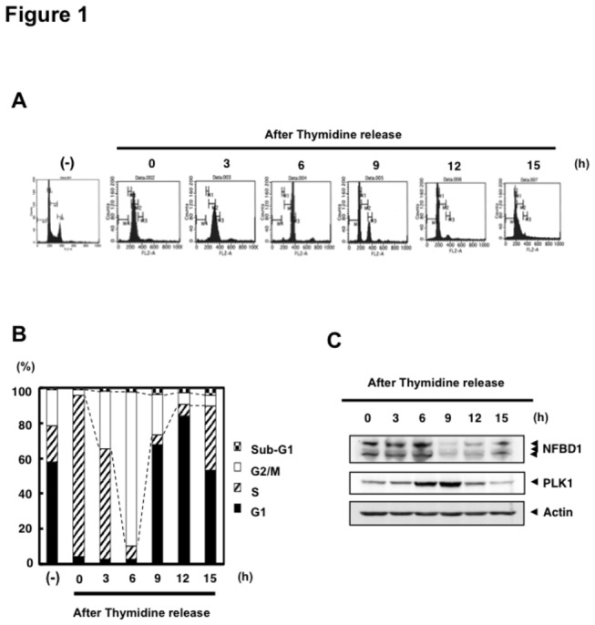
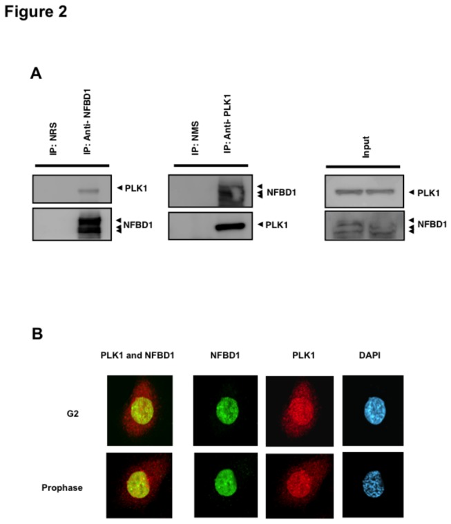
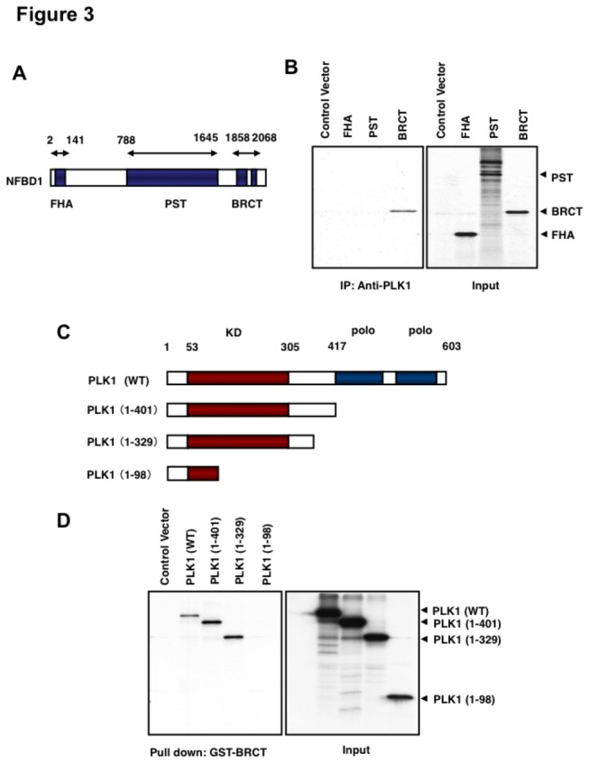
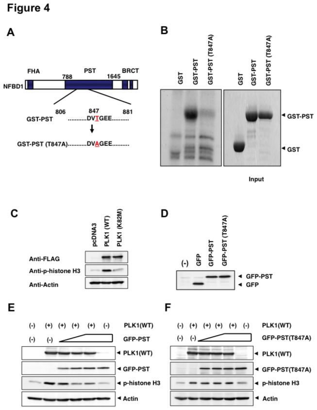
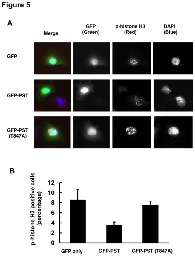
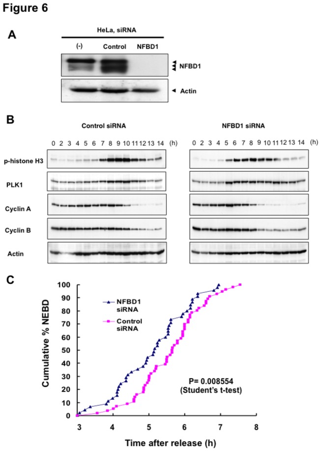
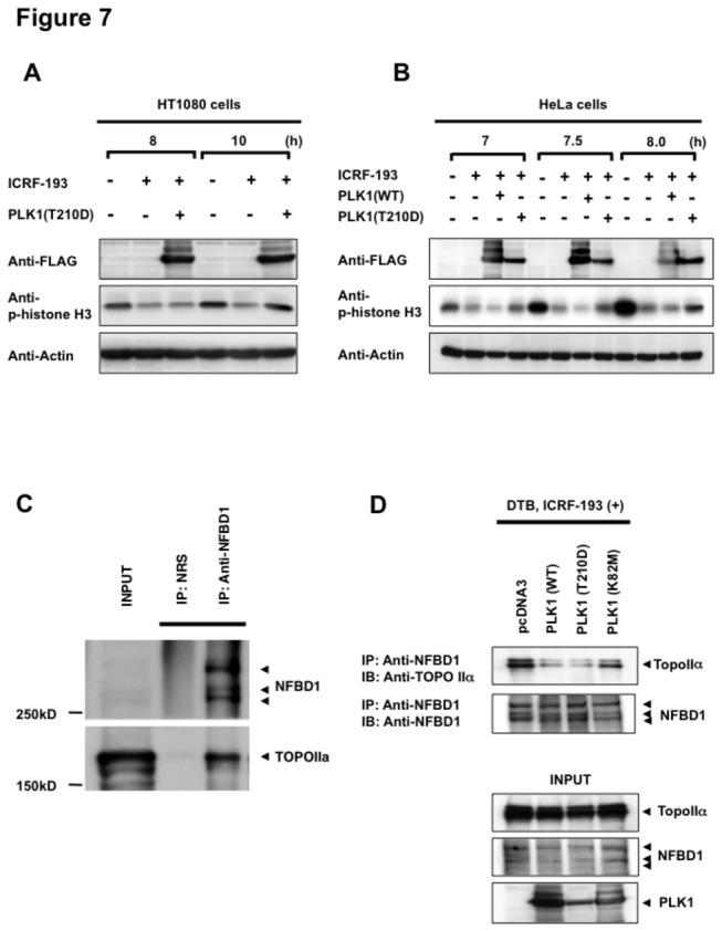
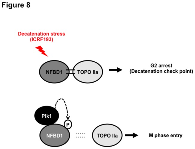
Although it has been established that nuclear factor with BRCT domain 1/ mediator of the DNA damage checkpoint protein 1 (NFBD1/MDC1) is closely involved in DNA damage response, its possible contribution to the regulation of cell- cycle progression is unclear. In the present study, we have found for the first time that NFBD1 is phosphorylated by polo-like kinase 1 (PLK1) and has an important role in G2/M transition. Both NFBD1 and PLK1 are co-expressed in cellular nuclei throughout G2/M transition, and binding assays demonstrated direct interaction between NFBD1 and PLK1. Indeed, in vitro kinase reactions revealed that the PST domain of NFBD1 contains a potential amino acid sequence (845-DVTGEE-850) targeted by PLK1. Furthermore, enforced expression of GFP-PST but not GFP-PST(T847A) where threonine at 847 was substituted by alanine inhibited the phosphorylation levels of histone H3, suggesting a defect of M phase entry. Because PLK1 has been implicated in promoting the G2/M transition, we reasoned that overexpressed PST might serve as a pseudosubstrate for PLK1 and thus interfere with phosphorylation of endogenous PLK1 substrates. Interestingly, siRNA-mediated knockdown of NFBD1 resulted in early M phase entry and accelerated M phase progression, raising the possibility that NFBD1 is a PLK1 substrate for regulating the G2/M transition. Moreover, the constitutive active form of PLK1(T210D) overcame the ICRF-193-induced decatenation checkpoint and inhibited the interaction between NFBD1 and topoisomerase IIα, but kinase-deficient PLK1 did not. Based on these observations, we propose that PLK1-mediated phosphorylation of NFBD1 is involved in the regulation of G2/M transition by recovering a decatenation checkpoint.


Conflict of interest statement

Competing Interests: The authors have declared that no competing interests exist.

Figures

Figure 1
Figure 1. NFBD1 and PLK1 are both expressed in the G2/M phase and form a protein complex.
(A) HeLa cells were double-thymidine blocked at the late G1 phase and then released into fresh medium. At the indicated times after release, cells were stained with propidium iodide (PI) and analyzed by FACS. (B) Schematic representation of cell-cycle distributions under the abovementioned experimental conditions. (C) Western blot analysis. At the indicated time after release, whole cell lysates were prepared and immunoblotted against anti-NFBD1 (first panel), anti-PLK1 (second panel), or anti-actin (third panel) antibody. Immunoblotting for actin is shown as a control for protein loading.
Figure 2
Figure 2. Co-immunoprecipitation and co-localization between NFBD1 and PLK1 in the G2/M phase.
(A) Immunoprecipitation analysis. HeLa cells were synchronized by the double-thymidine block regimen. Six hours after the second release, whole cell lysates prepared from HeLa cells were immunoprecipitated with normal rabbit serum or with a polyclonal anti-NFBD1 antibody followed by immunoblotting with a monoclonal anti-PLK1 antibody (left panel). Reciprocal experiments using normal mouse serum and a monoclonal anti-PLK1 antibody are shown in the middle panel. (B) Indirect immunofluorescence staining. HeLa cells were synchronized by the double-thymidine block regimen. Six hours after the second release, mitotic cells were simultaneously stained with polyclonal anti-NFBD1 (green) and monoclonal anti-PLK1 antibodies (red). Merged images (yellow) indicate the co-localization of NFBD1 with PLK1. Nuclear DNA was stained with DAPI (blue).
Figure 3
Figure 3. Mapping of the binding regions between NFBD1 and PLK1.
(A) A schematic drawing of the structure of NFBD1 is shown. Numbers indicate the amino acid positions relative to the first Met (+1). FHA, forkhead-associated; PST, proline/serine/threonine-rich; BRCT, BRCA1 carboxyl terminus. (B) In vitro binding assay. Whole cell lysates prepared from COS7 cells transfected with FLAG-Plk1 expression plasmid were incubated with 35S-labeled FHA, PST, or the BRCT domain of NFBD1. Bound materials were recovered by immunoprecipitation with an anti-PLK1 antibody and analyzed by SDS-PAGE followed by autoradiography (left panel). The right panel shows 1/10 loading. (C) The structure of wild-type Plk1 and various COOH-terminal deletion mutants of PLK1. KD, kinase domain; polo, polo box domain. (D) GST pull-down assay. 35S-labeled wild-type PLK1 or the indicated deletion mutants of PLK1 were incubated with GST-BRCT fusion protein. After the incubation, bound proteins were recovered on glutathione Sepharose beads and separated by SDS-PAGE followed by autoradiography (left panel). The right panel shows 1/10 loading.
Figure 4
Figure 4. PLK1-mediated phosphorylation of NFBD1 is essential for the M phase entry.
(A) Schematic representation of PST domains of wild-type NFBD1 and mutant form of NFBD1 termed GST-PST(T847A). (B) In vitro kinase reaction. GST, GST-PST, or GST-PST(T847A) were purified using glutathione Sepharose beads (right panel) and incubated with purified PLK1 in the presence of [γ-32P]ATP. The reaction mixtures were analyzed by SDS-PAGE followed by autoradiography (left panel). (C) Enforced expression of PLK1 but not the kinase-deficient mutant form of PLK1 [PLK1(K28M)] induces phospho-histone H3. HeLa cells were transiently transfected with the indicated expression plasmids. Forty-eight hours after transfection, whole cell lysates were prepared and immunoblotted with the indicated antibodies. (D) Expression of GFP-PST and GFP-PST(T847A). HeLa cells were transiently transfected with the expression plasmids. Forty-eight hours after transfection, whole cell lysates were prepared and immunoblotted with an anti-GFP antibody. (E and F) GFP-PST but not GFP-PST(T847A) inhibits the phosphorylation of histone H3. HeLa cells were transiently transfected with the expression plasmid for FLAG-PLK1 alone or FLAG-PLK1 and increasing amounts of GFP-PST (E) or GFP-PST(T847A) (F). Forty-eight hours after transfection, whole cell lysates were prepared and analyzed by immunoblotting with the indicated antibodies.
Figure 5
Figure 5. GFP-PST inhibits the phosphorylation of histone H3.
(A) Indirect immunofluorescence staining. HeLa cells were transiently transfected with the indicated expression plasmids. Forty-eight hours after transfection, cells were fixed and stained with polyclonal anti-phospho-histone H3 antibody (red). Nuclear DNA was stained with DAPI (blue). Representative photos demonstrate the initial H3 phosphorylation in pericentric heterochromatin during the late G2 phase. (B) The number of GFP-positive cells (green) with phosphorylated histone H3 was scored, and the percentage of phospho-histone H3-positive cells in each column represents the mean of three independent experiments.
Figure 6
Figure 6. Knockdown of NFBD1 accelerates G2/M progression.
(A) siRNA-mediated knockdown of NFBD1 results in a reduction of endogenous NFBD1. HeLa cells were synchronized by the double-thymidine block regimen and transfected with 10 nM of NFBD1 siRNA or control siRNA at the first release. At the time of the second release, whole cell lysates prepared from transfected cells were subjected to immunoblotting with anti-NFBD1 antibody (top panel). Western blotting for actin is shown as a control for protein loading (bottom panel). (B) Western blotting analysis of mitosis-related proteins. Whole cell lysates were prepared in NFBD1 siRNA transfected cells (right panel) or control siRNA transfected cells (left panel) at the indicated times after the second release and immunoblotted with anti-phospho-histone H3 antibody, anti-PLK1 antibody, anti-cyclin A antibody, anti-cyclin B antibody, or anti-actin antibody. (C) Cumulative percentages of NEBD-positive cells. GFP-cyclin B-expressing HeLa cells were synchronized and transfected with the indicated siRNAs. The times of NEBD were defined by the loss of a defined nuclear boundary and nuclear accumulation of cyclin B in the NFBD1 siRNA-transfected cells (n= 45) and control siRNA-transfected cells (n= 56). The significant difference was demonstrated by the Student’s t-test.
Figure 7
Figure 7. PLK1 inhibits the interaction between NFBD1 and TOPOII induced by the decatenation checkpoint.
(A and B) Decatenation checkpoint assay. HT1080 cells and HeLa cells were synchronized by the double-thymidine block regimen and transfected with PLK1(WT) or PLK1(T210D) at the first release. Transfected cells were treated with the indicated concentrations of ICRF-193 from the time of the second release. Whole cell lysates were prepared at the indicated times and subjected to immunoblotting with the indicated antibodies. (C) Interaction between NFBD1 and TOPOIIα. HeLa cells were synchronized and treated with 10 μM ICRF-193. Eight hours after the second release, whole cell lysates were prepared and subjected to anti-NFBD1 immunoprecipitation followed by immunoblotting with an anti-TOPOIIα antibody. Immunoprecipitation with NRS served as a negative control. The left panel shows 1/10 loading. (D) The effects of various kinase activities of PLK1 on the ICRF-193-induced interaction between NFBD1 and TOPOIIα. HeLa cells were synchronized and transfected with PLK1(WT), PLK1(T210D), PLk1(K82M), or control plasmid at the first release. Transfected cells were treated with ICRF-193 from the time of the second release. Whole cell lysates were prepared at 8 h after the second release and immunoprecipitated and subjected to immunoblotting with the indicated antibodies.
Figure 8
Figure 8. A model illustrating how NFBD1 contributes to the G2/M progression by PLK1-mediated phosphorylation.

References

    1. Goldberg M, Stucki M, Falck J, D'Amours D, Rahman D et al. (2003) MDC1 is required for the intra-S-phase DNA damage checkpoint. Nature 421: 952-956. doi:10.1038/nature01445. PubMed: 12607003. - DOI - PubMed
    1. Lou Z, Minter-Dykhouse K, Wu X, Chen J (2003) MDC1 is coupled to activated CHK2 in mammalian DNA damage response pathways. Nature 421: 957-961. doi:10.1038/nature01447. PubMed: 12607004. - DOI - PubMed
    1. Stewart GS, Wang B, Bignell CR, Taylor AM, Elledge SJ (2003) MDC1 is a mediator of the mammalian DNA damage checkpoint. Nature 421: 961-966. doi:10.1038/nature01446. PubMed: 12607005. - DOI - PubMed
    1. Lee MS, Edwards RA, Thede GL, Glover JN (2005) Structure of the BRCT repeat domain of MDC1 and its specificity for the free COOH-terminal end of the gamma-H2AX histone tail. J Biol Chem 280: 32053-32056. doi:10.1074/jbc.C500273200. PubMed: 16049003. - DOI - PubMed
    1. Stucki M, Clapperton JA, Mohammad D, Yaffe MB, Smerdon SJ et al. (2005) MDC1 directly binds phosphorylated histone H2AX to regulate cellular responses to DNA double-strand breaks. Cell 123: 1213-1226. doi:10.1016/j.cell.2005.09.038. PubMed: 16377563. - DOI - PubMed

Publication types

MeSH terms