. 2013 Dec 9;8(12):e82819.
doi: 10.1371/journal.pone.0082819. eCollection 2013.

Effects of vitamin A on in vitro maturation of pre-pubertal mouse spermatogonial stem cells

Affiliations

Effects of vitamin A on in vitro maturation of pre-pubertal mouse spermatogonial stem cells

Albanne Travers et al. PLoS One. .

Abstract

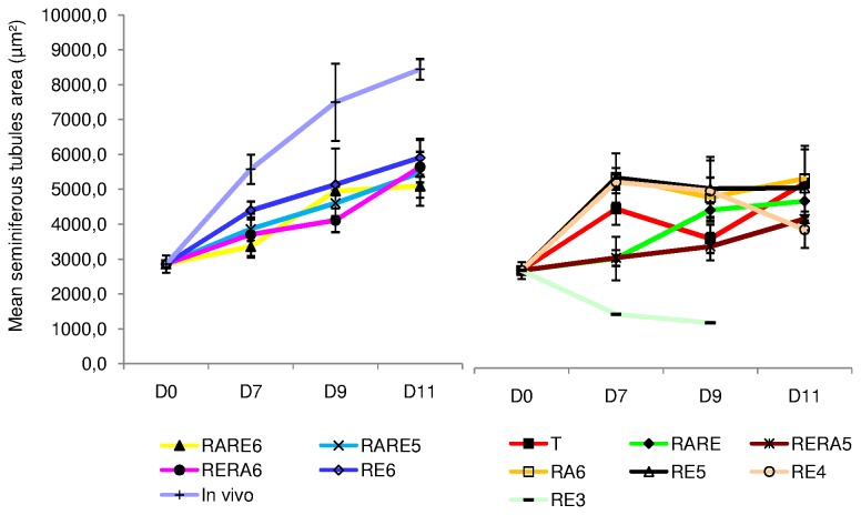
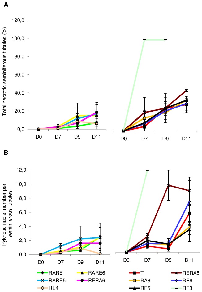
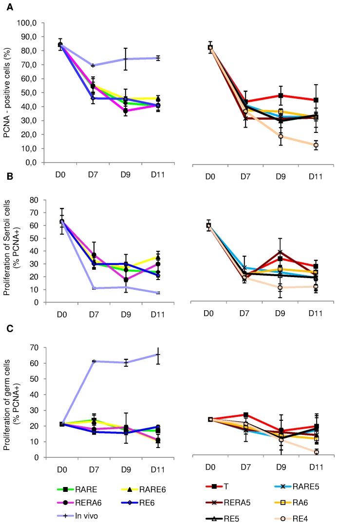
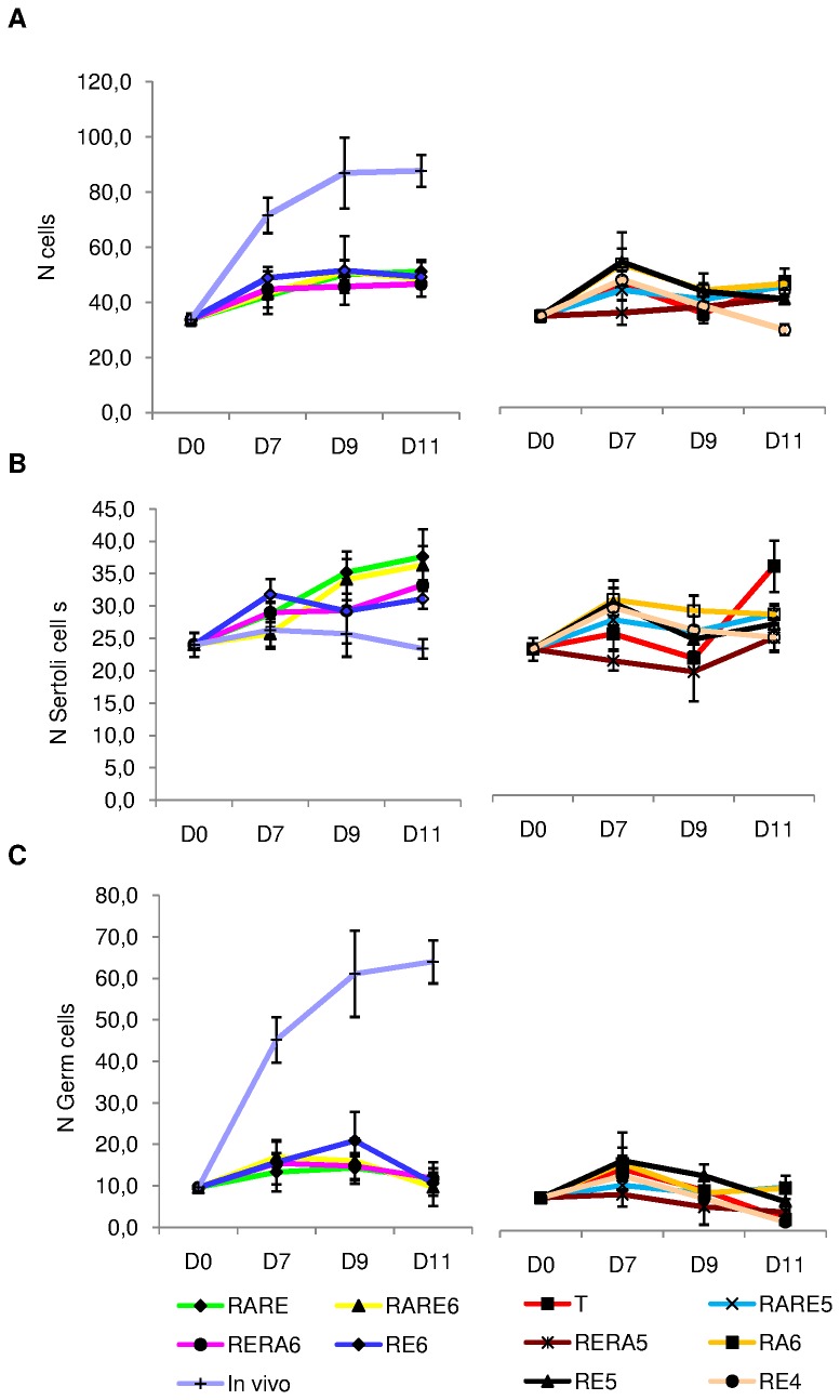
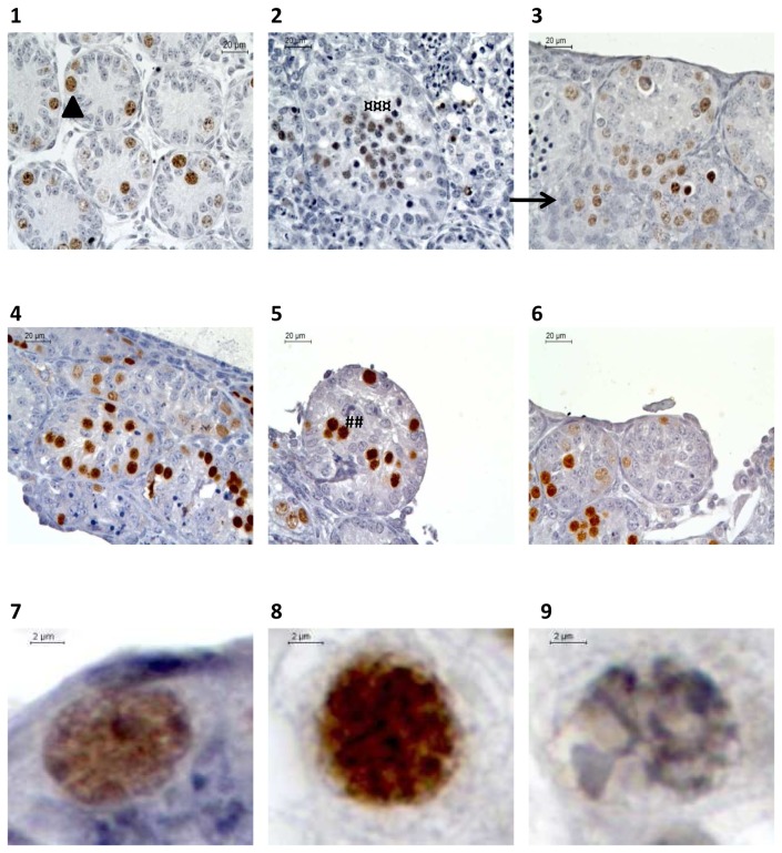
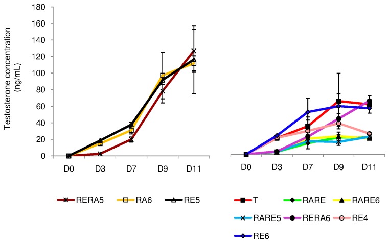
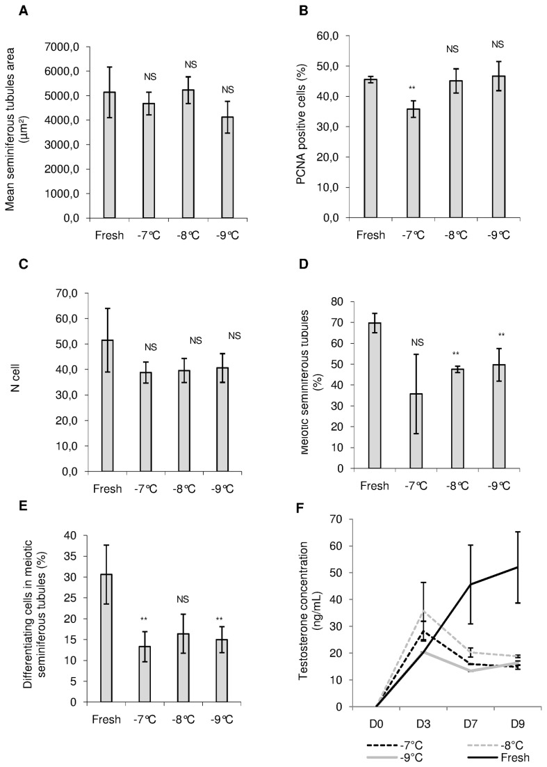
Testicular tissue cryopreservation is the only potential option for fertility preservation in pre-pubertal boys exposed to gonadotoxic treatment. Completion of spermatogenesis after in vitro maturation is one of the future uses of harvested testicular tissue. The purpose of the current study was to evaluate the effects of vitamin A on in vitro maturation of fresh and frozen-thawed mouse pre-pubertal spermatogonial stem cells in an organ culture system. Pre-pubertal CD1 mouse fresh testes were cultured for 7 (D7), 9 (D9) and 11 (D11) days using an organ culture system. Basal medium was supplemented with different concentrations of retinol (Re) or retinoic acid (RA) alone or in combination. Seminiferous tubule morphology (tubule diameter, intra-tubular cell type), intra-tubular cell death and proliferation (PCNA antibody) and testosterone level were assessed at D7, D9 and D11. Pre-pubertal mouse testicular tissue were frozen after a soaking temperature performed at -7 °C, -8 °C or -9 °C and after thawing, were cultured for 9 days, using the culture medium preserving the best fresh tissue functionality. Retinoic acid at 10(-6)M and retinol at 3.3.10(-7)M, as well as retinol 10(-6)M are favourable for seminiferous tubule growth, maintenance of intra-tubular cell proliferation and germ cell differentiation of fresh pre-pubertal mouse spermatogonia. Structural and functional integrity of frozen-thawed testicular tissue appeared to be well-preserved after soaking temperature at -8 °C, after 9 days of organotypic culture using 10(-6)M retinol. RA and Re can control in vitro germ cell proliferation and differentiation. Re at a concentration of 10(-6)M maintains intra-tubular cell proliferation and the ability of spermatogonia to initiate spermatogenesis in fresh and frozen pre-pubertal mouse testicular tissue using a soaking temperature at -8 °C. Our data suggested a possible human application for in vitro maturation of cryopreserved pre-pubertal testicular tissue.


Conflict of interest statement

Competing Interests: The authors have declared that no competing interests exist.

Figures

Figure 1
Figure 1. Seminiferous tubule growth (A) and intra-tubular necrosis (B) during organotypic culture of prepubertal mice testes under the different culture conditions assessed.
(A) Seminiferous tubule area was defined using software (red circle) and area was automatically generated (green rectangle). (B) The percentage of partially (arrow head) or totally (star) necrotic seminiferous tubules were assessed as well as number of pyknotic nuclei (head). (1-2-3) Organotypic culture of fresh immature mice testicular tissue after 0 (1) or 11 (2 and 3) days of culture with RE6 (2) or RERA5 (3) (4-5-6) Organotypic culture of frozen-thawed testicular tissue with stabilisation phase at –8°C (5) and -9°C (6) compared to fresh control organotypic culture (4) Footnotes: RE6: 10-6M retinol ; RERA5: 10-5M retinoic acid and 3.3.10-7M retinol
Figure 2
Figure 2. Seminiferous tubule area assessment in organotypic culture of fresh pre-pubertal mouse testicular tissue.
Seminiferous tubule area evolution after 0, 7, 9 and 11 days of organotypic culture on at least 30 cross-sectioned tubules after Tra98 immunostaining under the different culture conditions. The results are represented as the mean ± SEM with n=6. Footnotes: T: basal culture medium without retinoid; RARE: 3.3.10-7M RA and 3.3.10-7M RE; RARE6: 3.3.10-7M RA and 10-6M RE; RARE5: 3.3.10-7M RA and 10-5M RE; RERA6: 3.3.10-7M RE and 10-6M RA; RERA5: 3.3.10-7M RE and 10-5M RA; RA6: 10-6M RA; RE6: 10-6M RE; RE5: 10-5M RE; RE4: 10-4M RE; RE3: 10-3M RE; In vivo: In vivo control.
Figure 3
Figure 3. Cellular death assessment in organotypic culture of fresh pre-pubertal mouse testicular tissue.
Cellular death was assessed in organotypic culture at days 0, 7, 9 and 11 on 30 cross-sectioned seminiferous tubules after Tra98 immunolabelling under the different culture conditions tested. (A) Percentage of total pyknotic seminiferous tubules. (B) Number of pyknotic nuclei per seminiferous tubules. The results are represented as the mean ± SEM with n=6. Footnotes: T: basal culture medium without retinoid; RARE: 3.3.10-7M RA and 3.3.10-7M RE; RARE6: 3.3.10-7M RA and 10-6M RE; RARE5: 3.3.10-7M RA and 10-5M RE; RERA6: 3.3.10-7M RE and 10-6M RA; RERA5: 3.3.10-7M RE and 10-5M RA; RA6: 10-6M RA; RE6: 10-6M RE; RE5: 10-5M RE; RE4: 10-4M RE; In vivo: In vivo control. RA: Retinoic acid; RE: Retinol; D0: Day 0; D7: Day 7; D9: Day 9; D11: Day 11
Figure 4
Figure 4. Assessment of cellular proliferation after PCNA (Proliferating Cell Nuclear Antigen) and Tra98 immunodetection on serial sections of seminiferous tubules in organotypic culture of fresh and frozen-thawed pre-pubertal mouse testicular tissue.
(A) Cellular proliferation assessment for organotypic culture of fresh immature mice testicular tissue assessed after 0 (1 and 4) or 11 days (2,3,5,6) of culture with RE6 (2 and 5) or RERA5 (3 and 6) (B) Cellular proliferation assessment for organotypic culture of frozen-thawed testicular tissue with stabilisation phase at –8°C (2 and 5) and -9°C (3 and 6) compared to fresh control organotypic culture (1 and 4) 1 to 3 : PCNA immunodetection 4 to 6 : Tra98 iommunostaining ** : Germ cells PCNA positive ; ¤¤ : Sertoli cell PCNA positive ; ^^ : Sertoli cell PCNA negative Footnotes: RE6 : 10-6M retinol ; RERA5 : 10-5M retinoic acid and 3.3.10-7M retinol
Figure 5
Figure 5. PCNA (Proliferating Cell Nuclear Antigen) expression in organotypic culture of fresh pre-pubertal mouse testicular tissue.
PCNA expression was assessed in organotypic culture at days 0, 7, 9 and 11 on at least 500 intra-tubular cells under the different culture conditions tested. The results are represented as the mean ± SEM with n=6. (A) Percentage of total PCNA-positive cells. (B) Percentage of PCNA-positive Sertoli cells. (C) Percentage of PCNA-positive germ cells. FootnotesT: basal culture medium without retinoid; RARE: 3.3.10-7M RA and 3.3.10-7M RE; RARE6: 3.3.10-7M RA and 10-6M RE; RARE5: 3.3.10-7M RA and 10-5M RE; RERA6: 3.3.10-7M RE and 10-6M RA; RERA5: 3.3.10-7M RE and 10-5M RA; RA6: 10-6M RA; RE6: 10-6M RE; RE5: 10-5M RE; RE4: 10-4M RE; In vivo: In vivo control. RA: Retinoic acid; RE: Retinol; D0: Day 0; D7: Day 7; D9: Day 9; D11: Day 11
Figure 6
Figure 6. Cellular proliferation in organotypic culture of fresh pre-pubertal mouse testicular tissue on days 0, 7, 9 and 11.
Intratubular cell quantification (germ cells and Sertoli cells) was assessed after Tra98 immunodetection on 30 cross-sectioned seminiferous tubules for each tested concentration of retinoids. The results are represented as the mean ± SEM with n=6. (A) Total number of intratubular cells. (B) Number of Sertoli cells per seminiferous tubules. (C) Number of germ cells per seminiferous tubules. Footnotes: T: basal culture medium without retinoid; RARE: 3.3.10-7M RA and 3.3.10-7M RE; RARE6: 3.3.10-7M RA and 10-6M RE; RARE5: 3.3.10-7M RA and 10-5M RE; RERA6: 3.3.10-7M RE and 10-6M RA; RERA5: 3.3.10-7M RE and 10-5M RA; RA6: 10-6M RA; RE6: 10-6M RE; RE5: 10-5M RE; RE4: 10-4M RE; In vivo: In vivo control. RA: Retinoic acid; RE: Retinol; D0: Day 0; D7: Day 7; D9: Day 9; D11: Day 11
Figure 7
Figure 7. Histological evaluation of meiotic process evaluation after Tra98 immunostaining of organotypic culture of fresh and frozen-thawed pre-pubertal mice testicular tissue.
(1-2-3) Meiotic germ cells assessment for organotypic culture of fresh pre-pubertal mice testicular tissue at ×500 magnification. Fresh testicular tissue was culture for 0 (1) or 11 days (2 and 3) with RE6 (2) or RERA5 (3) (4-5-6) Meiotic germ cells assessment for organotypic culture of frozen-thawed (5 and 6) immature mice testicular tissue compared to fresh control (4) at ×500 magnification. Testicular tissue was frozen using a temperature stabilisation phase at –8°C (5) or –9°C (6) (7-8-9) Germ cells classification after Tra98 immunodtection at x1000 magnification. Intra-tubular cells were classified as spermatogonia (7, arrow head) (smooth spherical brown nuclei), leptotene/zygotene primary spermatocytes (8, ###) (irregular spherical brown nuclei with condensed chromatin) or pachytene primary spermatocytes (9, ¤¤¤) (irregular spherical brown nuclei with highly condensed chromatin). Sertoli cells was defined with a blue nucleus (head). Footnotes: RE6: 10-6M retinol ; RERA5: 10-5M retinoic acid and 3.3.10-7M retinol
Figure 8
Figure 8. Meiotic process in organotypic culture of fresh pre-pubertal mouse testicular tissue at days 0, 7, 9 and 11.
Meiotic germ cells assessment performed after Tra98 immunodetection on 30 cross-sectioned seminiferous for each tested concentration of retinoids. The results are represented as the mean ± SEM with n=6. (A) Percentage of seminiferous tubules in meiotic prophase (i.e containing leptotene, zygotene or pachytene primary spermatocytes). (B) Percentage of primary spermatocytes (i.e. leptotene, zygotene or pachytene) in meiotic seminiferous tubules. FootnotesT: basal culture medium without retinoid; RARE: 3.3.10-7M RA and 3.3.10-7M RE; RARE6: 3.3.10-7M RA and 10-6M RE; RARE5: 3.3.10-7M RA and 10-5M RE; RERA6: 3.3.10-7M RE and 10-6M RA; RERA5: 3.3.10-7M RE and 10-5M RA; RA6: 10-6M RA; RE6: 10-6M RE; RE5: 10-5M RE; RE4: 10-4M RE; In vivo: In vivo control. RA: Retinoic acid; RE: Retinol; D0: Day 0; D7: Day 7; D9: Day 9; D11: Day 11
Figure 9
Figure 9. Evolution of testosterone concentrations (ng/mL) in culture media collected between D0 and D11.
The results are represented as the mean ± SEM with n=6. Footnotes: T: basal culture medium without retinoid; RARE: 3.3.10-7M RA and 3.3.10-7M RE; RARE6: 3.3.10-7M RA and 10-6M RE; RARE5: 3.3.10-7M RA and 10-5M RE; RERA6: 3.3.10-7M RE and 10-6M RA; RERA5: 3.3.10-7M RE and 10-5M RA; RA6: 10-6M RA; RE6: 10-6M RE; RE5: 10-5M RE; RE4: 10-4M RE; In vivo: In vivo control. RA: Retinoic acid; RE: Retinol; D0: Day 0; D7: Day 7; D9: Day 9; D11: Day 11
Figure 10
Figure 10. Structural and functional assessment of frozen-thawed pre-pubertal mouse testicular tissue after 9 days of culture with 10-6M retinol.
Results were compared with fresh pre-pubertal testicular tissue cultured with the same conditions. Testicular tissue was cryopreserved using a controlled slow freezing protocol and a soaking temperature evaluated at -7°C, -8°C or -9°C. (A) Evolution of mean seminiferous tubule area on 30 cross-sectioned seminiferous tubules after Tra98 immunolabelling. (B) Cellular proliferation assessed by PCNA (Proliferating cell nuclear antigen) staining. (C) Quantification of intratubular cells after 9 days of culture using Tra98 immunodetection. (D) and (E) Meiosis assessment (i.e. leptotene, zygotene and pachytene spermatocytes I), after Tra98 immunostaining, using percentage of seminiferous tubules in meiotic prophase (D) or by assessment of percentage of primary spermatocytes (i.e. leptotene, zygotene or pachytene) in seminiferous tubules (E). (F) Steroidogenesis evolution. The results are represented as the mean ± SEM with n=6. NS : Not significant ; ** : p<0.01

References

    1. Dohle GR (2010) Male infertility in cancer patients: Review of the literature. Int J Urol 17: 327-331. doi:10.1111/j.1442-2042.2010.02484.x. PubMed: 20202000. - DOI - PubMed
    1. Schrader M, Müller M, Straub B, Miller K (2001) The impact of chemotherapy on male fertility: a survey of the biologic basis and clinical aspects. Reprod Toxicol 15: 611-617. doi:10.1016/S0890-6238(01)00182-4. PubMed: 11738514. - DOI - PubMed
    1. Tournaye H, Goossens E, Verheyen G, Frederickx V, De Block G et al. (2004) Preserving the reproductive potential of men and boys with cancer: current concepts and future prospects. Hum Reprod Update 10: 525-532. doi:10.1093/humupd/dmh038. PubMed: 15319377. - DOI - PubMed
    1. Menon S, Rives N, Mousset-Siméon N, Sibert L, Vannier JP et al. (2009) Fertility preservation in adolescent males: experience over 22 years at Rouen University Hospital. Hum. Reprod 24: 37-44. - PubMed
    1. Ginsberg JP, Carlson CA, Lin K, Hobbie WL, Wigo E et al. (2010) An experimental protocol for fertility preservation in prepubertal boys recently diagnosed with cancer: a report of acceptability and safety. Hum Reprod 25: 37-41. doi:10.1093/humrep/dep371. PubMed: 19861330. - DOI - PMC - PubMed

Publication types