. 2013 Dec 11;8(12):e83172.
doi: 10.1371/journal.pone.0083172. eCollection 2013.

Alveolar macrophage innate response to Mycobacterium immunogenum, the etiological agent of hypersensitivity pneumonitis: role of JNK and p38 MAPK pathways

Affiliations

Alveolar macrophage innate response to Mycobacterium immunogenum, the etiological agent of hypersensitivity pneumonitis: role of JNK and p38 MAPK pathways

Harish Chandra et al. PLoS One. .

Abstract

Mycobacterium immunogenum is an emerging pathogen of the immune-mediated lung disease hypersensitivity pneumonitis (HP) reported in machinists occupationally exposed to contaminated metal working fluid (MWF). However, the mechanism of its interaction with the host lung is unclear. Considering that alveolar macrophages play a central role in host defense in the exposed lung, understanding their interaction with the pathogen could provide initial insights into the underlying immunopathogenesis events and mechanisms. In the current study, M. immunogenum 700506, a predominant genotype isolated from HP-linked fluids, was shown to multiply intracellularly, induce proinflammatory mediators (TNF-α, IL-1α, IL-1β, IL-6, GM-CSF, NO) and cause cytotoxicity/cell death in the cultured murine alveolar macrophage cell line MH-S in a dose- and time-dependent manner. The responses were detected as early as 3h post-infection. Comparison of this and four additional genotypes of M. immunogenum (MJY-3, MJY-4, MJY-12, MJY-14) using an effective dose-time combination (100 MOI for 24h) showed these macrophage responses in the following order (albeit with some variations for individual response indicators). Inflammatory: MJY-3 ≥ 700506 > MJY-4 ≥ MJY-14 ≥ MJY-12; Cytotoxic: 700506 ≥ MJY-3 > MJY-4 ≥ MJY-12 ≥ MJY-14. In general, 700506 and MJY-3 showed a more aggressive response than other genotypes. Chemical blocking of either p38 or JNK inhibited the induction of proinflammatory mediators (cytokines, NO) by 700506. However, the cellular responses showed a somewhat opposite effect. This is the first report on M. immunogenum interactions with alveolar macrophages and on the identification of JNK- and p38- mediated signaling and its role in mediating the proinflammatory responses during these interactions.


Conflict of interest statement

Competing Interests: The authors have declared that no competing interests exist.

Figures

Figure 1
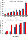
Figure 1. Induction of cellular responses (in terms of LDH release and cell death) in the MH-S cells after 24h exposure to M. immunogenum 700506.
A. Effect of varying the infection dose. B. Effect of time course after exposure to a selected infection dose (100 MOI). Values presented as means ± standard deviations are from three independent experiments; each experiment was done in triplicate. Asterisk (*) indicates statistically significant (P≤0.05) difference as compared to the vehicle control.
Figure 2
Figure 2. Determination of intracellular replication of M. immunogenum 700506 in alveolar macrophages (MH-S cells).
A. Cells infected with bacteria for 1 hour were made free of extracellular bacteria by repeated (3x) washing followed by gentamycin treatment as described under Materials and Methods section and their intracellular bacterial load was monitored for 72 hours based on CFU analysis. B. Inactivation of extracellular bacteria in gentamycin treatment. Log CFU values presented as means ± standard deviations are from three independent experiments; each experiment was done in triplicate. Asterisk (**) indicates statistically significant (P≤0.01) difference as compared to the values for other time points. C& D. Transmission electron microscopy (TEM) images of uninfected (C) and MI-infected (D) cells at a magnification of 30000X. Black arrows indicate the intracellular M. immunogenum cells. Abbreviations: Gm (gentamycin), Mi (M. immunogenum), Nuc (nucleus), Vac (vacuole), and Mito (mitochondrion), respectively.
Figure 3
Figure 3. Induction of nitric oxide (NO) production (measured as nitrite) in MH-S cells after 24h exposure to M. immunogenum 700506.
A. Effect of varying the infection dose. B. Time course of NO production after exposure to a selected dose (100 MOI). Values are presented as means ± standard deviations calculated based on the data obtained from three independent experiments; each experiment was done in triplicate. Asterisk (*) indicates statistically significant (P≤0.05) difference as compared to the vehicle control.
Figure 4
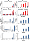
Figure 4. Induction of cytokine response in MH-S cells after 24h post-exposure to M. immunogenum 700506.
A. Effect of varying the infection dose on the expression of different cytokines in MH-S cells at 24h post-infection (a) IL-1α (b) IL-1β, (c) TNF-α, (d) IL-6, (e) GM-CSF. B. Time course of expression of different cytokines in MH-S cells exposed to an infection dose of 100 MOI (a) IL-1α (b) IL-1β, (c) TNF-α, (d) IL-6, (e) GM-CSF. Values are presented as means ± standard deviations calculated based on the data obtained from three independent experiments; each experiment was done in triplicate. A statistically significant (P≤0.05) value as compared to vehicle control is indicated by an asterisk (*).
Figure 5
Figure 5. Comparison of M. immunogenum genotypes 700506, MJY-3, MJY-4, MJY-12 and MJY-14 for induction of cellular response in terms of LDH release and cell viability loss in MH-S cells.
The cells were infected at a 100 MOI dose for 24h. Values are presented as means ± standard deviations calculated based on the data obtained from three independent experiments; each experiment was done in triplicate. Asterisk (*) indicates statistically significant (P≤0.05) difference from the Vehicle-treated group and pound sign (#) indicates statistically significant (P≤0.05) difference from the 700506-treated group.
Figure 6
Figure 6. Comparison of M. immunogenum genotypes 700506, MJY-3, MJY-4, MJY-12 and MJY-14 for induction of proinflammatory mediators (cytokines/NO) in MH-S cells.
The cells were infected at a 100 MOI dose for 24h. A & B. Levels of proinflammatory cytokines IL-1α and IL-1β in both culture supernatant and cell lysate. C, D, & E. Levels of TNF- α, IL-6 and GM-CSF in cell culture supernatant. F. Level of NO measured as nitrite in cell culture supernatant. Asterisk (*) and pound sign (#) indicate statistically significant (P≤0.05) difference from the vehicle-treated and 700506-treated groups, respectively. Values are presented as means ± standard deviations calculated based on the data obtained from three independent experiments; each experiment was done in triplicate.
Figure 7
Figure 7. Activation of MAPKs in alveolar macrophages on infection with M. immunogenum 700506.
A. p38 immunoblot. B. SAPK/JNK immunoblot. Activation was assessed in terms of upregulation of total MAPK expression levels and increase in phosphorylated MAPK. Densitometric analysis of the Western blots was done using the NIH’s Image J software. Lanes 1-8 represent vehicle control (VC), 0.001, 0.01, 0.1, 1, 10, 100 and 1000 MOI, respectively. Details on the antibodies for total- and phospho- MAPKs and β-actin are described under the Materials and Methods section. Values are presented as means ± standard deviations calculated based on the data obtained from three independent experiments; each experiment was done in triplicate. Asterisk (*) indicates statistically significant (P≤0.05) difference as compared to the vehicle control.
Figure 8
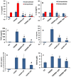
Figure 8. Effect of p38 inhibitor (SB202190) and JNK inhibitor (SP600125) on expression of proinflammatory mediators (cytokines/NO) in MH-S cells infected with M. immunogenum 700506.
A & B. Levels of proinflammatory cytokines IL-1α and IL-1β in cell culture supernatant and lysate. C, D, & E. Levels of TNF-α, IL-6, and GM-CSF in cell culture supernatant. F. Levels of NO production (measured as nitrite). Values are presented as means ± standard deviations calculated based on the data obtained from three independent experiments; each experiment was done in triplicate. Asterisk (*) and pound sign (#) indicate statistically significant (P≤0.05) difference from the vehicle control and 700506 (no inhibitor) control, respectively.
Figure 9
Figure 9. Effect of MAPK inhibitors on cellular responses (in terms of Cell viability and LDH release) in MH-S cells infected with M. immunogenum 700506.
A. Demonstration of the effect of the p38 inhibitor (SB202190) and JNK inhibitor (SP600125), each at 20 µM concentrations, in comparison with the no-inhibitor control (positive control) and vehicle control (negative control). Asterisk (*) and pound sign (#) indicate statistically significant (P≤0.05) difference from the vehicle control and 700506 (no inhibitor) control, respectively. B. Dose-dependent effect of p38 inhibitor (SB202190) and JNK inhibitor (SP600125) on the cellular responses using 20 µM and 40 µM concentrations. Asterisks (**) indicate statistically significant (P≤0.001) difference between different concentrations of inhibitor. Values are presented as means ± standard deviations calculated based on the data obtained from three independent experiments; each experiment was done in triplicate.

References

    1. Rosenman KD (2009) Asthma, hypersensitivity pneumonitis and other respiratory diseases caused by metalworking fluids. Curr Opin Allergy Clin Immunol 9: 97–102. - PubMed
    1. Falkinham JO (2003) The changing pattern of nontuberculous mycobacterial disease. Can J Infect Dis 14: 281–286. - PMC - PubMed
    1. Beckett W, Kallay M, Sood A, Zuo Z, Milton D (2005) Hypersensitivity pneumonitis associated with environmental mycobacteria. Environ Health Perspect 113: 767–770. - PMC - PubMed
    1. Shelton BG, Flanders WD, Morris GK (1999) Mycobacterium sp. as a possible cause of hypersensitivity pneumonitis in machine workers. Emerg Infect Dis 5: 270–273. - PMC - PubMed
    1. Wilson RW, Steingrube VA, Böttger EC, Springer B, Brown-Elliott BA, et al. (2001) Mycobacterium immunogenum sp. nov., a novel species related to Mycobacterium abscessus and associated with clinical disease, pseudo-outbreaks and contaminated metalworking fluids: an international cooperative study on mycobacterial taxonomy. Int J Syst Evol Microbiol 51: 1751–64. - PubMed

Publication types

MeSH terms