. 2013 Dec 12;8(12):e83584.
doi: 10.1371/journal.pone.0083584. eCollection 2013.

Functional cooperation between vitamin D receptor and Runx2 in vitamin D-induced vascular calcification

Affiliations

Functional cooperation between vitamin D receptor and Runx2 in vitamin D-induced vascular calcification

Min-Su Han et al. PLoS One. .

Abstract

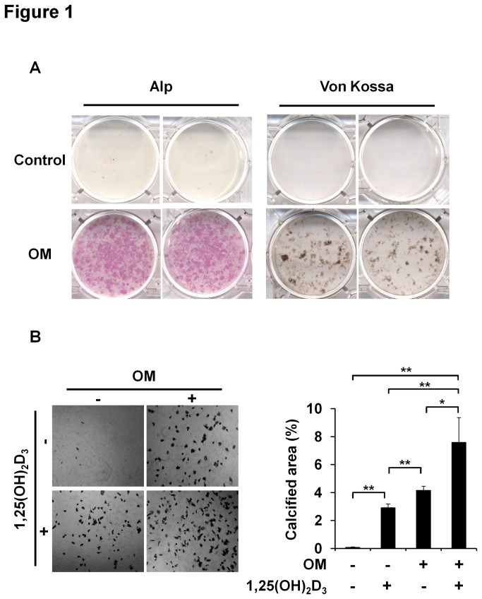
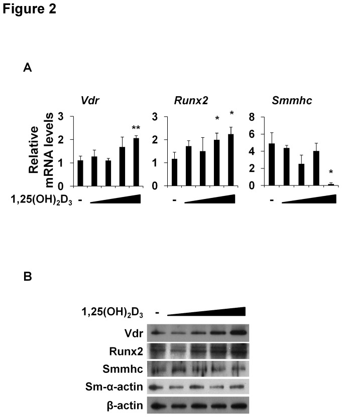
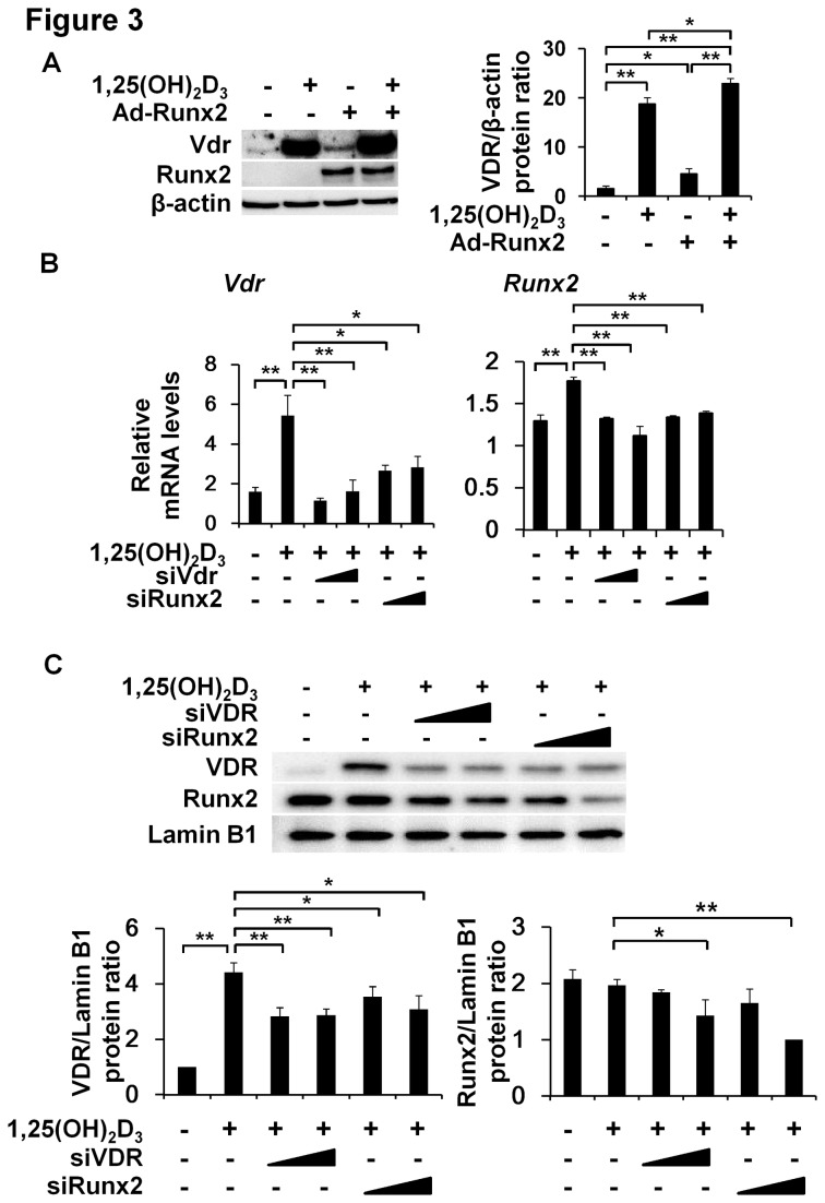
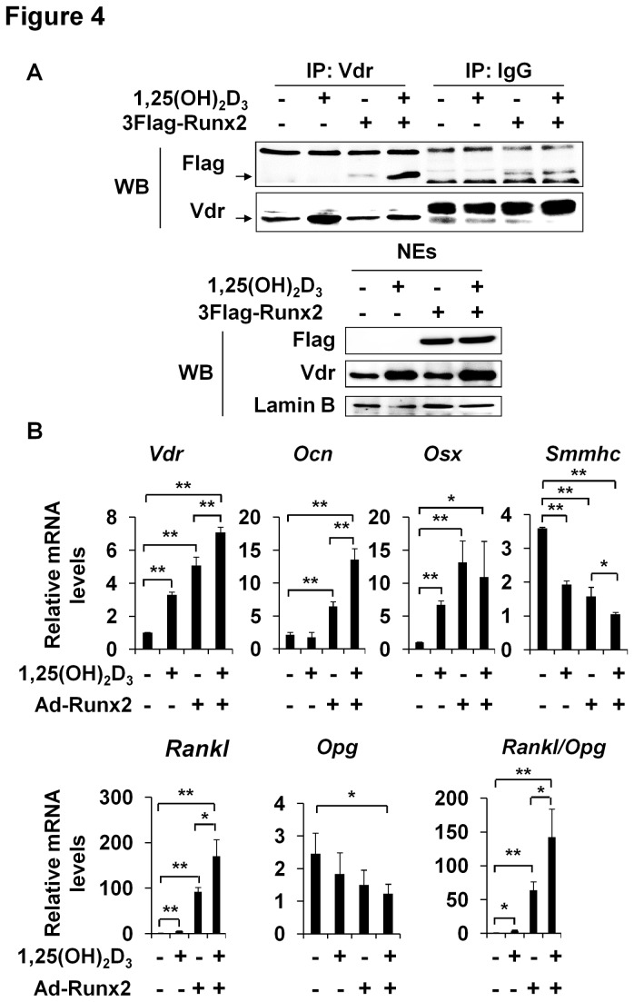
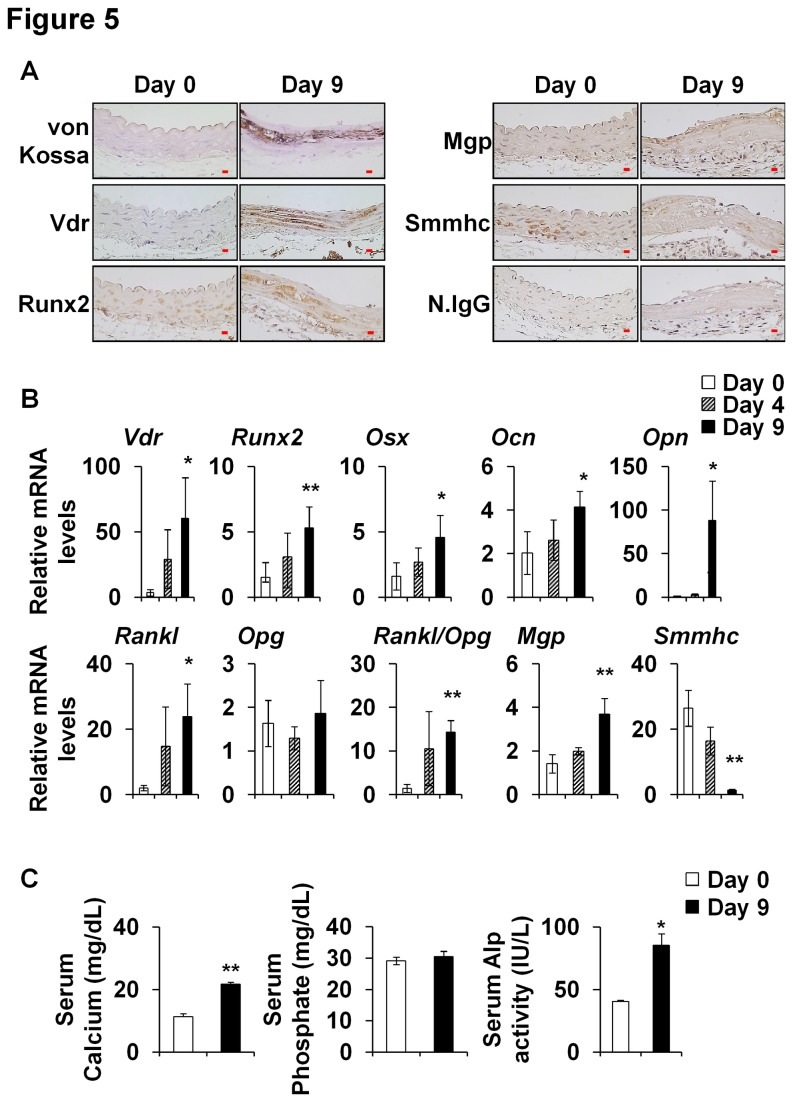
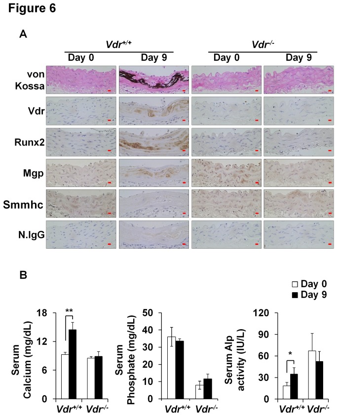
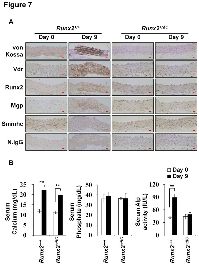
The transdifferentiation of vascular smooth muscle cells (VSMCs) into osteoblast-like cells has been implicated in the context of vascular calcification. We investigated the roles of vitamin D receptor (Vdr) and runt-related transcription factor 2 (Runx2) in the osteoblastic differentiation of VSMCs in response to vitamin D3 using in vitro VSMCs cultures and in vivo in Vdr knockout (Vdr(-/-)) and Runx2 carboxy-terminus truncated heterozygous (Runx2(+/ΔC)) mice. Treatment of VSMCs with active vitamin D3 promoted matrix mineral deposition, and increased the expressions of Vdr, Runx2, and of osteoblastic genes but decreased the expression of smooth muscle myosin heavy chain in primary VSMCs cultures. Immunoprecipitation experiments suggested an interaction between Vdr and Runx2. Furthermore, silencing Vdr or Runx2 attenuated the procalcific effects of vitamin D3. Functional cooperation between Vdr and Runx2 in vascular calcification was also confirmed in in vivo mouse models. Vascular calcification induced by high-dose vitamin D3 was completely inhibited in Vdr(-/-) or Runx2(+/ΔC) mice, despite elevated levels of serum calcium or alkaline phosphatase. Collectively, these findings suggest that functional cooperation between Vdr and Runx2 is necessary for vascular calcification in response to vitamin D3.


Conflict of interest statement

Competing Interests: The authors have declared that no competing interests exist.

Figures

Figure 1
Figure 1. Effects of 1,25(OH)2D3 on VSMC calcification.
(A) Primary mouse VSMCs were cultured in osteogenic medium (OM) or non-osteogenic control medium (Control). Alkaline phosphatase (Alp) and von Kossa staining were performed on culture day 21. (B) VSMCs were cultured with or without 1,25(OH)2D3 (10-7 mol/l) and von Kossa stained on day 21. Results are the means ± SD of three separate experiments (Right panel). Statistical analysis was performed using the unpaired Student’s t-test. *P<0.05 or **P<0.01 versus the untreated condition.
Figure 2
Figure 2. Effects of 1,25(OH)2D3on the expressions of Vdr and Runx2.
(A) Primary cultured mouse VSMCs were treated with 1,25(OH)2D3 (0, 10-10,10-9,10-8, or 10-7 mol/l) for 24 hours. Relative mRNA levels of Vdr, Runx2, and Smmhc were measured by real-time RT-PCR. (B) Protein levels of Vdr, Runx2, Smmhc, and Sm-α-actin were measured in total cell lysates by immunoblotting after treatment with 1,25(OH)2D3for 24 hours. β-Actin was used as an internal control. Statistical analysis was performed using the unpaired Student’s t-test. *P<0.05 or **P<0.01 versus the untreated condition.
Figure 3
Figure 3. Reciprocal regulation of Vdr and Runx2 by 1,25(OH)2D3.
(A) Rat VSMCs were infected with adenovirus expressing LacZ (Ad-LacZ) or Runx2 (Ad-Runx2) vectors (50 moi) for 4 hours and then treated with or without 1,25(OH)2D3 (1x10⁻⁷ mol/l) for 42 hours. The expression levels of Vdr and Runx2 were detected by immunoblotting. (B) Sub-confluent mouse VSMCs were seeded in 6-well plates, cultured overnight, and transfected with siRNAs (25 and 50 nmol/l) for Vdr or Runx2 mRNAs and then treated with 1,25(OH)2D3 (except the control) for 24 hours. Total RNAs were subjected to real-time RT PCR. (C) Mouse VSMCs cultured for 48 hours were analyzed for Vdr and Runx2 protein levels. Lamin B1 was used as internal control. Results are the means ± SD of three independent experiments. Statistical analysis was analyzed using the unpaired Student’s t-test. *P<0.05; **P<0.01.
Figure 4
Figure 4. Physical interaction between Vdr and Runx2 and the expressions of bone related genes.
(A) HEK293 cells were transfected with Flag-Runx2 and treated with 1,25(OH)2D3 (1 x 10⁻⁷ mol/l) for 24 hours. Nuclear extracts (NEs) were immunoprecipitated with anti-Vdr antibody and then immunoblotted with anti-Flag or anti-Vdr antibodies (upper panel). NEs were also immunoblotted with anti-Flag, anti-Vdr, and anti-Lamin B antibodies (lower panel). (B) Rat VSMCs were infected with Ad-LacZ or Ad-Runx2 vectors (at 50 moi) for 4 hours and then treated with or without 1,25(OH)2D3 (1 x 10⁻⁷ mol/l) for 42 hours. Relative mRNA levels of Vdr, Ocn, Osx, Rankl, Opg, and Smmhc were determined by real-time RT-PCR. Data are the means ± SDs of three separate experiments. Statistical analysis was analyzed using the unpaired Student’s t-test. *P<0.05 ; **P<0.01.
Figure 5
Figure 5. Vitamin D3-induced VC and the expression of bone related genes in the aortas of wild type mice.
After administering high-dose vitamin D3 (6 x 10⁵ IU/kg of body weight) injection to wild type mice as described in Materials and Methods, aortas were obtained at day 0 (baseline), day 4 and day 9 and subjected to immunohistochemical or real-time RT-PCR assays. (A) Calcium deposition was observed by von Kossa staining. Vdr, Runx2, and Mgp protein levels were increased but Smmhc was decreased in calcified regions. Original magnification, X400 (Scale bar=10 μm). Mice (n=6-8) per group. Normal IgG (N.Igg) was used as an internal control. (B) The mRNA levels of Vdr, Runx2, Osx, Ocn, Opn, Rankl, Opg, Mgp, and Smmhc were measured by real-time RT-PCR. Data are group means ± SD (n=6-8/group). (C) Serum calcium and phosphate levels, and Alp activities were measured. Data are group means ± SD of experimental groups (n=6-8). Statistical analysis was performed using the unpaired Student’s t-test. *P<0.05; **P<0.01.
Figure 6
Figure 6. Protection from vitamin D3-induced VC in Vdr-/- mice.
Sixteen-week-old wild type (Vdr+/+) and Vdr-/- mice were injected with vitamin D3 (8 x 10⁵ IU/kg of body weight). (A) Regions of calcification were detected by von Kossa and immunohistochemical staining. Vdr, Runx2, and Mgp protein levels were elevated in calcified regions in Vdr+/+ but not in Vdr-/- mice. Original magnification, X400 (Scale bar=10 μm). Control, n=5; vitamin D3, n=5. N.IgG was used as an internal control. (B) Serum calcium, phosphate, and Alp levels were also analyzed. Data are group means ± SDs (n=5/group). Statistical analysis was performed using the unpaired Student’s t-test. *P<0.05; **P<0.01.
Figure 7
Figure 7. Protection from vitamin D3-induced VC in Runx2 +/ΔC mice.
Eight-week-old Runx2+/+ and Runx2 +/ΔC mice were injected with vitamin D3 (6 x 10⁵ IU/kg of body weight). (A) Calcified regions were detected by von Kossa staining. The protein levels of Vdr, Runx2, Mgp, and Smmhc in these regions were assessed by immunohistochemical staining. Original magnification, X400 (Scale bar=10 μm). Experimental groups (n=5-8). N.IgG was used as an internal control. (B) Serum calcium, phosphate, and Alp levels were also measured. Data are group means ± SDs (n=5-8/group). Statistical analysis was analyzed using the unpaired Student’s t-test. *P<0.05; **P<0.01.

References

    1. Vattikuti R, Towler DA (2004) Osteogenic regulation of vascular calcification: an early perspective. Am J Physiol Endocrinol Metab 286: E686-E696. doi:10.1152/ajpendo.00552.2003. PubMed: 15102615. - DOI - PubMed
    1. Mody N, Tintut Y, Radcliff K, Demer LL (2003) Vascular calcification and its relation to bone calcification: possible underlying mechanisms. J Nucl Cardiol 10: 177-183. doi:10.1067/mnc.2003.0012. PubMed: 12673183. - DOI - PubMed
    1. Collin-Osdoby P (2004) Regulation of vascular calcification by osteoclast regulatory factors RANKL and osteoprotegerin. Circ Res 95: 1046-1057. doi:10.1161/01.RES.0000149165.99974.12. PubMed: 15564564. - DOI - PubMed
    1. Shao JS, Cheng SL, Sadhu J, Towler DA (2010) Inflammation and the osteogenic regulation of vascular calcification: a review and perspective. Hypertension 55: 579-592. doi:10.1161/HYPERTENSIONAHA.109.134205. PubMed: 20101002. - DOI - PMC - PubMed
    1. Khosla S (2011) The bone and beyond: a shift in calcium. Nat Med 17: 430-431. doi:10.1038/nm0411-430. PubMed: 21475237. - DOI - PubMed

Publication types

MeSH terms

LinkOut - more resources