

Psoriasis and cardiovascular risk factors: increased serum myeloperoxidase and corresponding immunocellular overexpression by Cd11b(+) CD68(+) macrophages in skin lesions
- PMID: 24349618
- PMCID: PMC3853421
Psoriasis and cardiovascular risk factors: increased serum myeloperoxidase and corresponding immunocellular overexpression by Cd11b(+) CD68(+) macrophages in skin lesions
Abstract
Background: Recent studies report independent associations between psoriasis, cardiovascular (CV) events and risk factors. Blood Myeloperoxidase (MPO) from activated myeloid cells is associated with CV risk mainly through lipid oxidation, induction of endothelial dysfunction and release of IL-12 from macrophages.
Objectives: To elucidate associations between psoriasis and conventional CV risk factors.
Methods: We performed a cross-sectional study of 100 psoriasis patients and 53 controls, group matched on age, gender and body mass index, to assess levels of MPO in serum, as well as immunohistochemical staining from psoriasis skin lesions, psoriasis uninvolved skin, and normal skin.
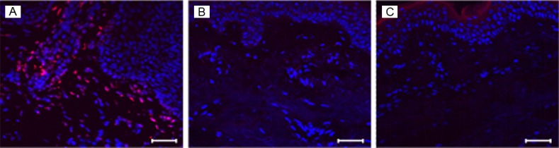
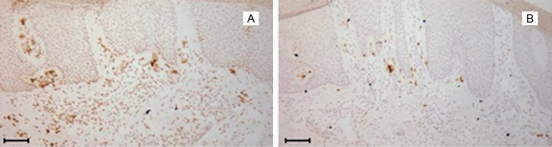
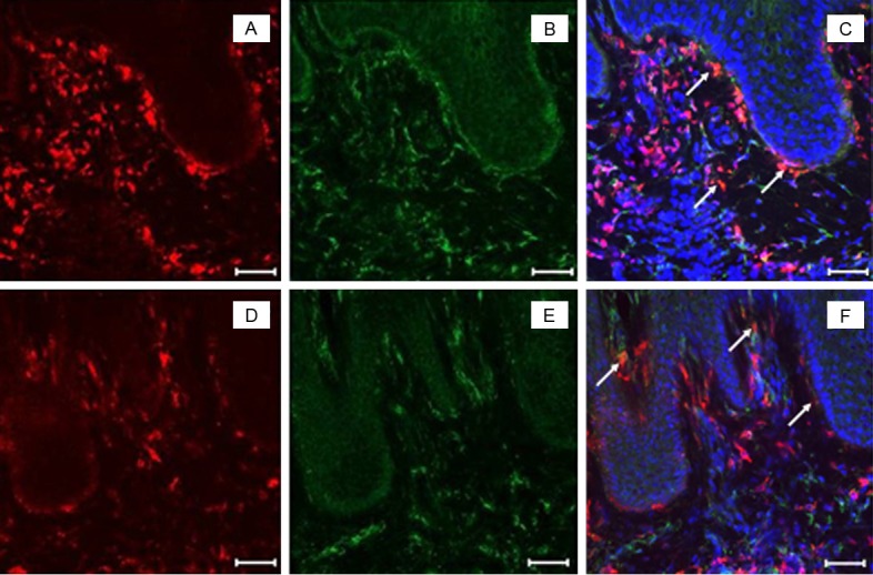
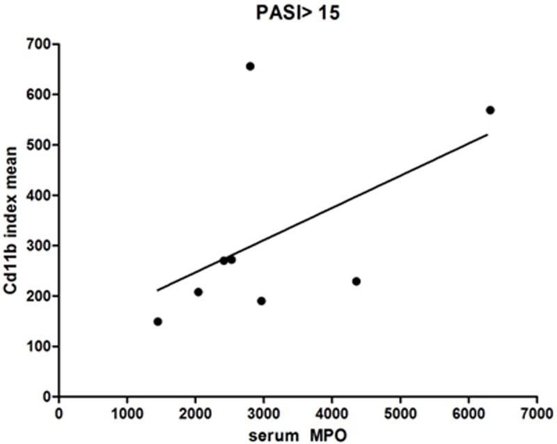
Results: Although the groups did not differ on waist circumference, glucose, cholesterol, triglycerides, creatinine or personal history of CV events, psoriasis patients had significantly higher waist-to-hip ratios, blood pressures, proportion of current smokers, and lower high density lipoprotein level than controls. Serum MPO level was elevated 2.5 fold (P<0.001) in psoriasis patients, even after adjusting for the CV risk factors on which the groups differed. MPO did correlate with coronary artery calcification, carotid plaque, carotid intima media thickness and flow mediated dilation, but did not correlate with psoriasis severity. However, MPO was highly expressed in lesional psoriatic skin and colocalized predominantly with CD45(+) CD11b(+) leukocytes. CD11b(+) cell density correlated with circulation MPO levels.
Conclusion: Lesional skin CD11b(+) leukocytes activated to generate MPO may contribute to serum levels of MPO. Lesional CD11b(+) cell activity may be an alternative measure of disease burden to PASI that underlies the MPO biomarker for systemic inflammation related to Cardiovascular Disease.
Keywords: Myeloperoxidase; cardiovascular disease; immunofluorescence; immunohistochemistry; psoriasis.
Figures


References
-
- NPF. About psoriasis: statistics. 2009. National Psoriasis Foundation.
-
- Gelfand JM, Neimann AL, Shin DB, Wang X, Margolis DJ, Troxel AB. Risk of myocardial infarction in patients with psoriasis. JAMA. 2006;296:1735–41. - PubMed
-
- del Rincon ID, Williams K, Stern MP, Freeman GL, Escalante A. High incidence of cardiovascular events in a rheumatoid arthritis cohort not explained by traditional cardiac risk factors. Arthritis Rheum. 2001;44:2737–45. - PubMed
-
- Rhew EY, Ramsey-Goldman R. Premature atherosclerotic disease in systemic lupus erythematosus--role of inflammatory mechanisms. Autoimmun Rev. 2006;5:101–5. - PubMed
Grants and funding
LinkOut - more resources
Full Text Sources
Other Literature Sources
Medical
Research Materials
Miscellaneous