

Screening and early diagnosis of osteoporosis through X-ray and ultrasound based techniques
- PMID: 24349644
- PMCID: PMC3856332
- DOI: 10.4329/wjr.v5.i11.398
Screening and early diagnosis of osteoporosis through X-ray and ultrasound based techniques
Abstract
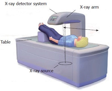
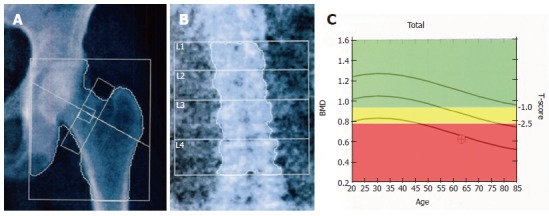
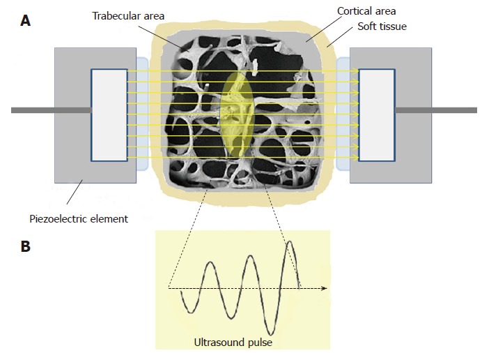
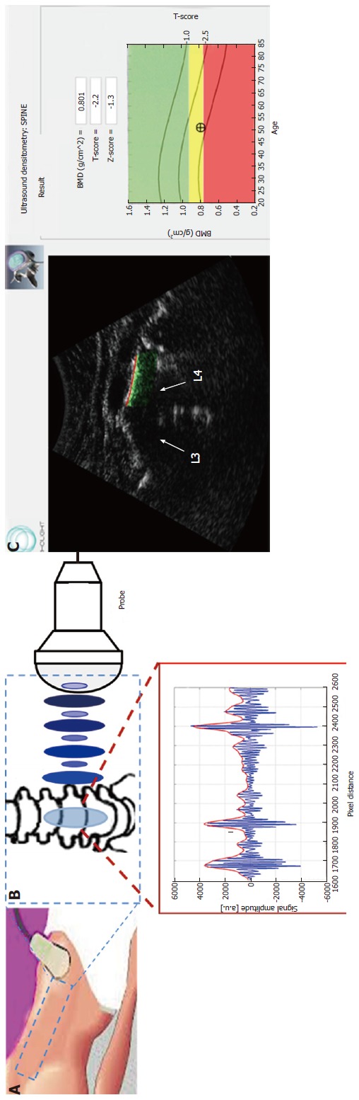
Effective prevention and management of osteoporosis would require suitable methods for population screenings and early diagnosis. Current clinically-available diagnostic methods are mainly based on the use of either X-rays or ultrasound (US). All X-ray based methods provide a measure of bone mineral density (BMD), but it has been demonstrated that other structural aspects of the bone are important in determining fracture risk, such as mechanical features and elastic properties, which cannot be assessed using densitometric techniques. Among the most commonly used techniques, dual X-ray absorptiometry (DXA) is considered the current "gold standard" for osteoporosis diagnosis and fracture risk prediction. Unfortunately, as other X-ray based techniques, DXA has specific limitations (e.g., use of ionizing radiation, large size of the equipment, high costs, limited availability) that hinder its application for population screenings and primary care diagnosis. This has resulted in an increasing interest in developing reliable pre-screening tools for osteoporosis such as quantitative ultrasound (QUS) scanners, which do not involve ionizing radiation exposure and represent a cheaper solution exploiting portable and widely available devices. Furthermore, the usefulness of QUS techniques in fracture risk prediction has been proven and, with the last developments, they are also becoming a more and more reliable approach for assessing bone quality. However, the US assessment of osteoporosis is currently used only as a pre-screening tool, requiring a subsequent diagnosis confirmation by means of a DXA evaluation. Here we illustrate the state of art in the early diagnosis of this "silent disease" and show up recent advances for its prevention and improved management through early diagnosis.
Keywords: Bone mineral density; Diagnosis of osteoporosis; Peripheral sites; Quantitative ultrasound; Screening techniques; X-ray based methods.
Figures

References
-
- Kanis JA, Burlet N, Cooper C, Delmas PD, Reginster JY, Borgstrom F, Rizzoli R; European Society for Clinical and Economic Aspects of Osteoporosis and Osteoarthritis (ESCEO) European guidance for the diagnosis and management of osteoporosis in postmenopausal women. Osteoporos Int. 2008;19:399–428. - PMC - PubMed
-
- Albanese CV, De Terlizzi F, Passariello R. Quantitative ultrasound of the phalanges and DXA of the lumbar spine and proximal femur in evaluating the risk of osteoporotic vertebral fracture in postmenopausal women. Radiol Med. 2011;116:92–101. - PubMed
-
- Ensrud KE, Thompson DE, Cauley JA, Nevitt MC, Kado DM, Hochberg MC, Santora AC, Black DM. Prevalent vertebral deformities predict mortality and hospitalization in older women with low bone mass. Fracture Intervention Trial Research Group. J Am Geriatr Soc. 2000;48:241–249. - PubMed
-
- Cooper C. The crippling consequences of fractures and their impact on quality of life. Am J Med. 1997;103:12S–17S; discussion 17S-19S. - PubMed
-
- Kanis JA. Diagnosis of osteoporosis and assessment of fracture risk. Lancet. 2002;359:1929–1936. - PubMed
Publication types
LinkOut - more resources
Full Text Sources
Other Literature Sources
