

doi: 10.1155/2013/152528.
Epub 2013 Sep 19.
Clinical and imaging findings of true hemifacial hyperplasia
Affiliations
- PMID: 24349801
- PMCID: PMC3856155
- DOI: 10.1155/2013/152528
Item in Clipboard
Clinical and imaging findings of true hemifacial hyperplasia
Case Rep Dent.
2013.
Abstract
Congenital hemifacial hyperplasia is a rare developmental disorder of unknown etiology, characterized by a marked unilateral facial asymmetry. It involves the hard (bones and teeth) and soft tissues of the face. We report an interesting case of true hemifacial hyperplasia in a 25-year-old male highlighting the clinical and computed tomography imaging findings.
Figures
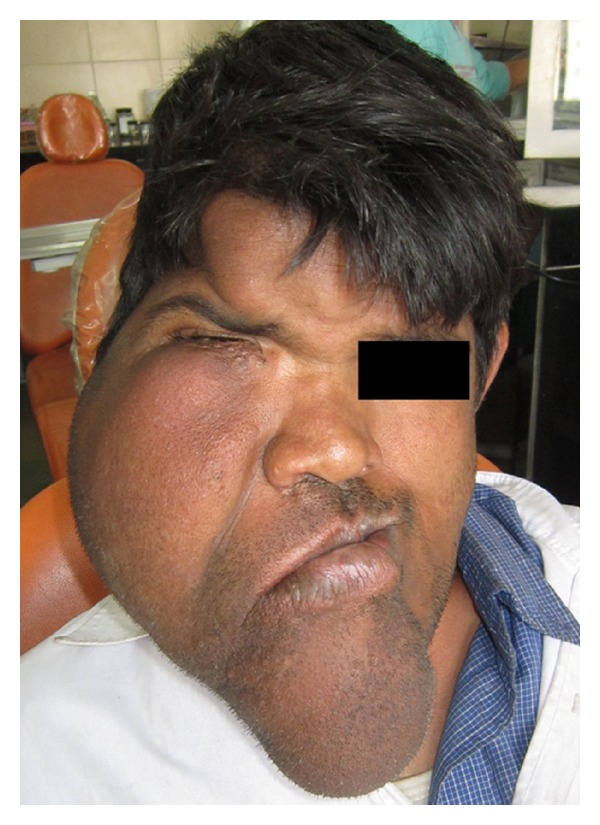
Unilateral enlargement of right side of the face. Note the asymmetry of the forehead, cheeks, nose, lips, chin, and the closed eye.
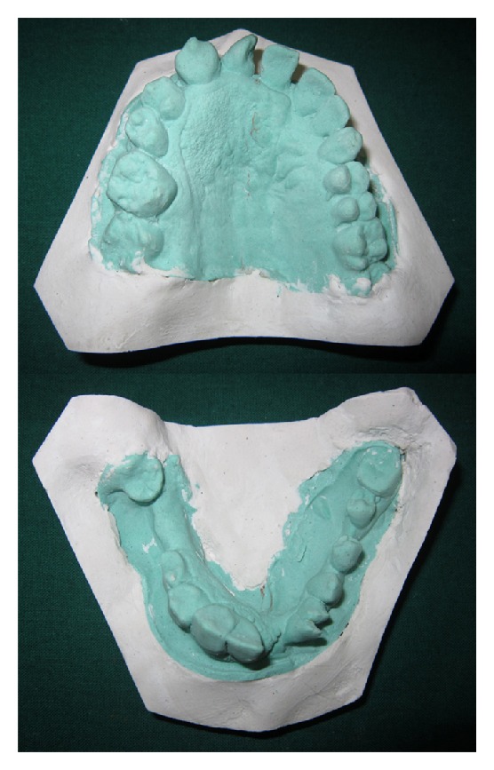
Occlusal view of the maxillary and mandibular dental casts showing macrodontia of the right side, midline shift to the left, along with granular surface of the right palatal mucosa.
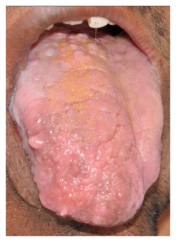
Intraoral photograph showing enlargement of right side of the tongue with enlarged papillae.
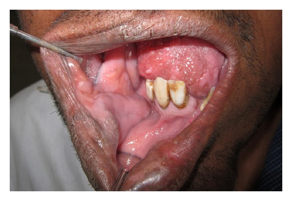
Intraoral photograph showing velvety buccal mucosa hanging in pendulous folds and granular gingival surface along with macrodontic permanent right lateral incisor and canine.
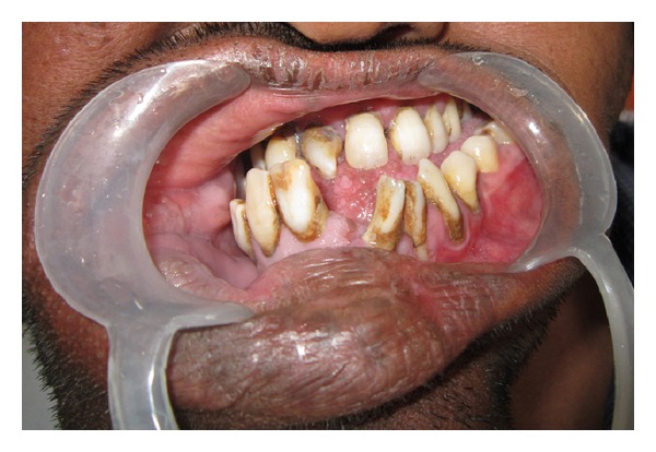
Intraoral photograph showing major variability between crown sizes of the teeth on the right and the left side with generalised crossbite relationship.
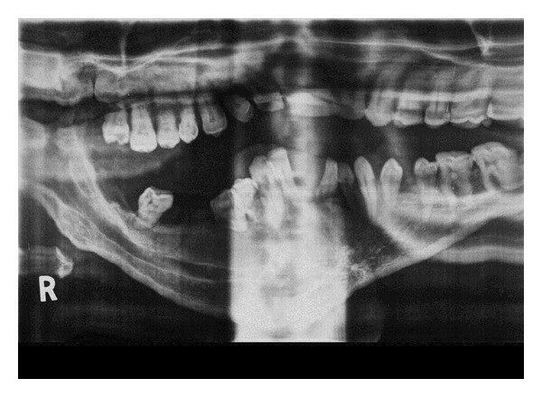
Panoramic radiograph showing widening of the right inferior alveolar canal.
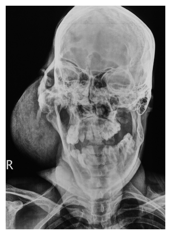
Posteroanterior view of the skull showing appreciable generalized bony and soft tissue enlargement of the right face.
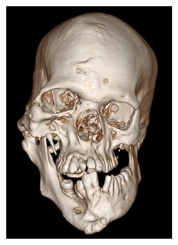
3D volume rendered CT scan showing enlarged mandible, maxilla, and zygoma, with enlarged right mental foramen and teeth of the right side.
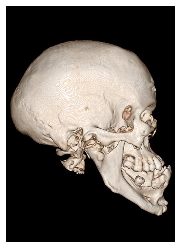
Lateral view of 3D volume rendered CT scan showing enlarged right condyle, coronoid process, mandibular body, and zygoma.
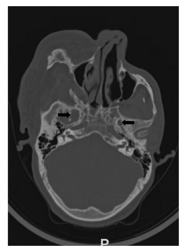
Axial CT scan with bone reconstructions showing widened foramen ovale (right side-arrow, left side-arrowhead).
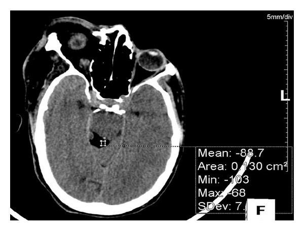
Axial CT scan showing a lipoma in the right quadrigeminal cistern.

Axial CT scan showing lipomatous enlargement of the soft tissue on right side of face including buccal region, lips, tongue, soft palate, and right parotid gland (asterix).
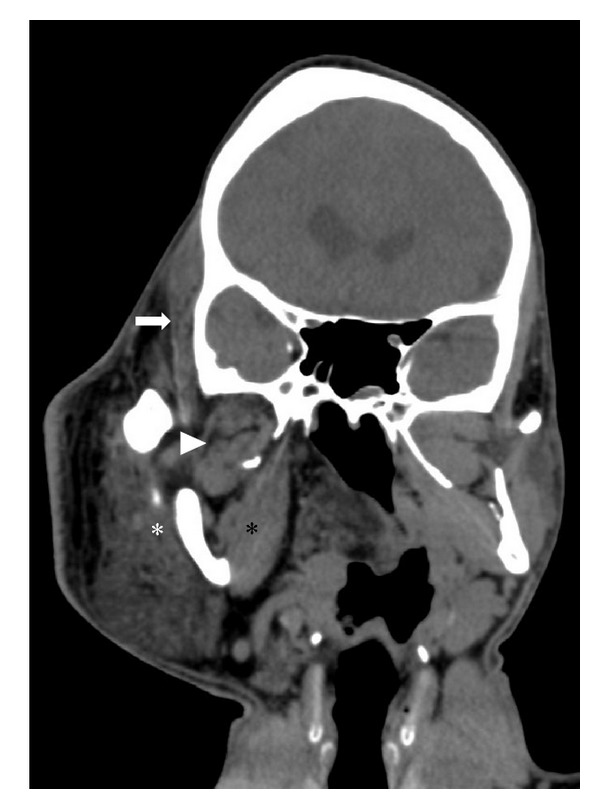
Coronal CT scan revealing hyperplasia of the right mandibular condyle, medial pterygoid (black asterisk), lateral pterygoid (arrowhead), masseter (white asterisk), and temporalis muscle (white arrow).
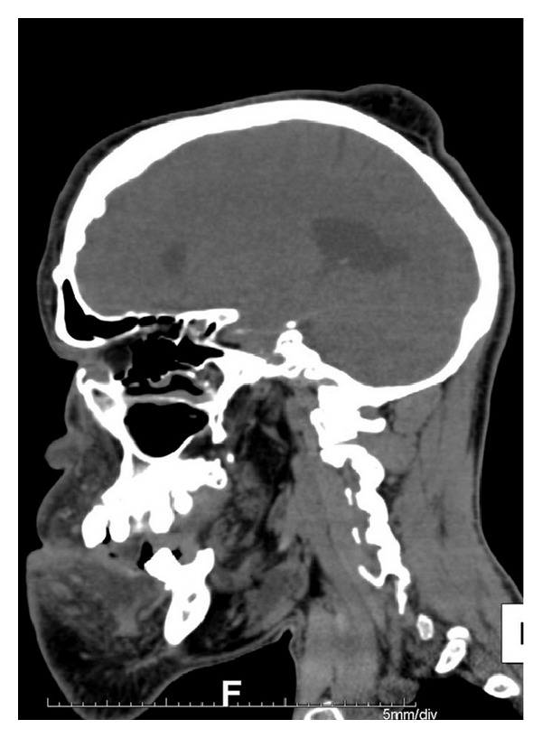
Sagital CT scan showing soft tissue swelling in right high parietal region.
References
-
- Meckel JF. Ueber die seitliche Asymmetric im tierischen Korper. In: Halle R, editor. Anatomische Physiologische Beobachtungen und Untersuchungen. 1822. p. p. 147.
-
- Kottmeier HL, Wagner HL. Über Hemihypertrophia und Hemiatrophia corporis totalis nebst spontane Extremitätengangräne bei Säuglingen im Anschluss zu einem ungewöhnlichen. Fall Acta Paediatrica. 1938;20(4):530–543.
-
- Rowe NH. Hemifacial hypertrophy—review of the literature and addition of four cases. Oral Surgery, Oral Medicine, Oral Pathology. 1962;15(5):572–587. - PubMed
-
- Pollock RA, Haskell Newman M, Burdi AR, Condit DP. Congenital hemifacial hyperplasia: an embryologic hypothesis and case report. Cleft Palate Journal. 1985;22(3):173–184. - PubMed
-
- Nayak R, Baliga MS. Crossed hemifacial hyperplasia: a diagnostic dilemma. Journal of Indian Society of Pedodontics & Preventive Dentistry. 2007;25(1):39–42. - PubMed
LinkOut - more resources
Full Text Sources
Other Literature Sources
