

Value of transesophageal echocardiography (TEE) guidance in minimally invasive mitral valve surgery
- PMID: 24349984
- PMCID: PMC3857007
- DOI: 10.3978/j.issn.2225-319X.2013.10.09
Value of transesophageal echocardiography (TEE) guidance in minimally invasive mitral valve surgery
Abstract
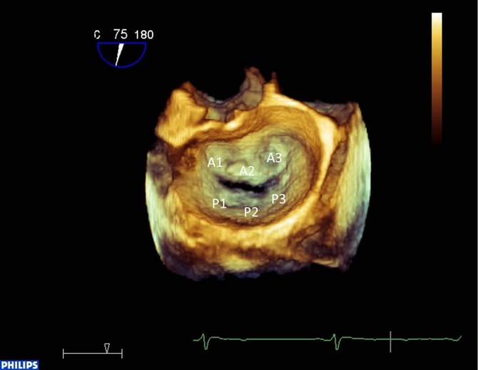
The role of intraoperative transesophageal echocardiography (TEE) has increased tremendously since its first use in 1979. Today intraoperative TEE is a class I indication for surgical mitral valve reconstruction for evaluation of mitral valve pathology, graduation of mitral regurgitation and detection of potential risk factors as well as post-repair assessment. Real-time three-dimensional TEE offers anatomical visualization of the mitral valve apparatus, fundamental for virtual surgical planning of proper annuloplasty ring size. As minimally invasive and even off-pump techniques for mitral valve repair become more popular, image guidance by intraoperative TEE will play an essential role.
Keywords: Intraoperative transesophageal echocardiography (TEE); image guidance; minimally invasive; mitral valve repair; real time three dimensional TEE (RT 3D TEE).
Figures


References
-
- Matsumoto M, Oka Y, Lin YT, et al. Transesophageal echocardiography; for assessing ventricular performance. N Y State J Med 1979;79:19-21 - PubMed
-
- Lancellotti P, Moura L, Pierard LA, et al. European association of echocardiography recommendations for the assessment of valvular regurgitation. Part 2: mitral and tricuspid regurgitation (native valve disease). Eur J Echocardiogr 2010;11:307-32 - PubMed
-
- Joint Task Force on the Management of Valvular Heart Disease of the European Society of Cardiology (ESC) , European Association for Cardio-Thoracic Surgery (EACTS), Vahanian A, et al. Guidelines on the management of valvular heart disease (version 2012). Eur Heart J 2012;33:2451-96 - PubMed
-
- Shanewise JS, Cheung AT, Aronson S, et al. ASE/SCA guidelines for performing a comprehensive intraoperative multiplane transesophageal echocardiography examination: recommendations of the American Society of Echocardiography Council for Intraoperative Echocardiography and the Society of Cardiovascular Anesthesiologists Task Force for Certification in Perioperative Transesophageal Echocardiography. Anesth Analg 1999;89:870-84 - PubMed
-
- Eltzschig HK, Rosenberger P, Loffler M, et al. Impact of intraoperative transesophageal echocardiography on surgical decisions in 12,566 patients undergoing cardiac surgery. Ann Thorac Surg 2008;85:845-52 - PubMed
LinkOut - more resources
Full Text Sources
Other Literature Sources
Research Materials