Comment
. 2013 Nov 5;110(45):E4178-82.
doi: 10.1073/pnas.1316666110.

Reply to Subramaniam, van Heel, and Henderson: Validity of the cryo-electron microscopy structures of the HIV-1 envelope glycoprotein complex

Comment

Reply to Subramaniam, van Heel, and Henderson: Validity of the cryo-electron microscopy structures of the HIV-1 envelope glycoprotein complex

Youdong Mao et al. Proc Natl Acad Sci U S A. .
No abstract available


Conflict of interest statement

The authors declare no conflict of interest.

Figures
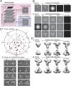
Fig. 1.
Fig. 1.
Validity of the HIV-1 Env(-)ΔCT structure. (A) In the dual-target function approach, particles of Env(-)ΔCT trimers are picked by fast local correlation (target function 1) with a low-resolution template. The picked particles are verified by ML alignment, which uses a log-likelihood function (target function 2). Even when the fast local correlation aligns pure noise images to produce an unaligned average image resembling the particle-picking template (reference bias), the ML alignment evaluates the picked particles by optimizing a different target function and removes the reference-biased artifactual feature (6). (B) ML alignment is resistant to bias from the starting reference and from the particle-picking template. The head of Einstein starting reference is rapidly removed by a few iterations of ML refinement of the average of 4,485 pure-noise images (panel 1). In the experiments shown in panel 2, a single projection of the HIV-1 envelope glycoprotein trimer was used to pick 4,485 “particles” from 200 pure noise micrographs of 4,096 × 4,096 pixels. The picked particles were subjected to ML alignment, using a starting reference of a pure noise image randomly chosen from the particle set (Top), a Gaussian circle (Middle), or the average of the picked particles without any further alignment (Bottom). This starting reference for ML optimization is shown in the first column. Each row shows the history of the ML-aligned averages at the indicated iterations of optimization, ending with the converged average in the far right column. In no case did the particle-picking template appear in the converged ML-aligned averages. (C) Tilt-pair raw cryo-EM images of the Env(-)ΔCT trimer were collected at experimental tilt angles of 0° and 10° and a tilt axis of 270° (panel 2) with a dose of ∼20 electron/Å2 in each exposure at 80 kV. The tilt-pair parameter plot is shown in panel 1, with each tilt pair of particles represented by a point. The radial value indicates the calculated tilt angle, whereas the azimuthal angle indicates the tilt axis. The cluster is indicated by a red circle, whose center is located at the mean tilt angle and axis. (D) Two RELION reconstructions were carried out on two independent datasets of the Env(-)ΔCT trimer. In panel 1, a 100-Å tetrahedron-like shape was used as a starting reference to refine the Env(-)ΔCT structure with a 9,993-particle dataset; the refinement converged at ∼20 Å. In panel 2, a low-pass-filtered 60-Å reference was used as a starting reference to refine the structure with a dataset of 124,478 particles; the refinement converged at 10 Å. No mask was used in the RELION refinement. Both RELION reconstructions are compatible with the published 11- and 6-Å Env(-)ΔCT structures (4, 5).
Fig. 2.
Fig. 2.
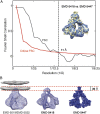
Comparison of cryo-EM density maps. (A) The FSC between the 11-Å map (EMD-5418) (4) and the 6-Å map (EMD-5447) (5) of the Env(-)ΔCT trimer is shown, with the critical FSC calculated from the two maps indicated by the red curve. The FSC curve crosses the critical FSC at ∼11 Å. The alignment of EMD-5418 (blue) and EMD-5447 (yellow) is shown in the Inset. The two maps are consistent, with more detailed features in the 6-Å map, as expected. (B) The map (EMD-5019) derived by cryo-electron tomography of the native, mature HIV-1 virion spike (19) is aligned with the 11- and 6-Å Env(-)ΔCT maps (EMD-5418 and EMD-5447, respectively) (4, 5). The reported location of the lipid bilayer membrane (EMD-5022) (19) with respect to the tomogram is shown as a gray mesh, with a 30-Å hydrophobic portion of the membrane indicated by the broken red lines. Except for differences in resolution, the three maps are closely related.

Comment on

References

    1. Subramaniam S. Structure of trimeric HIV-1 envelope glycoproteins. Proc Natl Acad Sci USA. 2013;110:E4172–E4174. - PMC - PubMed
    1. van Heel M. Finding trimeric HIV-1 envelope glycoproteins in random noise. Proc Natl Acad Sci USA. 2013;110:E4175–E4177. - PMC - PubMed
    1. Henderson R. Avoiding the pitfalls of single particle cryoEM: Einstein from noise. Proc Natl Acad Sci USA. 2013;110:18037–18041. - PMC - PubMed
    1. Mao Y, et al. Subunit organization of the membrane-bound HIV-1 envelope glycoprotein trimer. Nat Struct Mol Biol. 2012;19(9):893–899. - PMC - PubMed
    1. Mao Y, et al. Molecular architecture of the uncleaved HIV-1 envelope glycoprotein trimer. Proc Natl Acad Sci USA. 2013;110(30):12438–12443. - PMC - PubMed

MeSH terms

LinkOut - more resources