Review
. 2014 Nov 1;21(13):1926-42.
doi: 10.1089/ars.2013.5785. Epub 2014 Feb 19.

Role of reactive oxygen species in neonatal pulmonary vascular disease

Affiliations
Review

Role of reactive oxygen species in neonatal pulmonary vascular disease

Stephen Wedgwood et al. Antioxid Redox Signal. .

Abstract

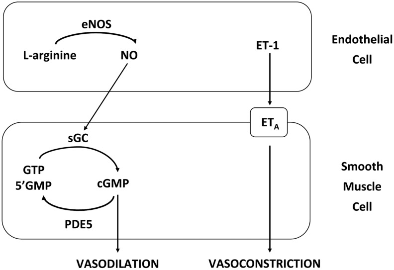
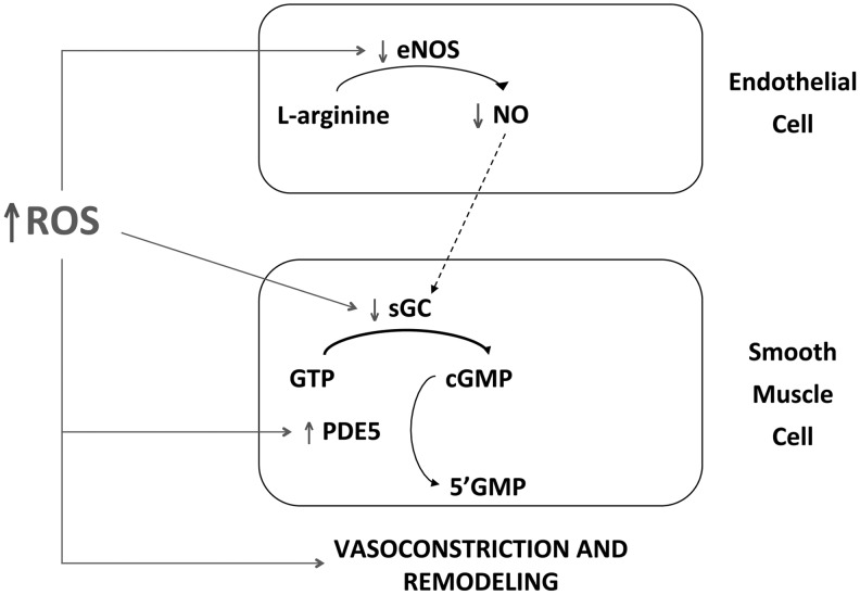
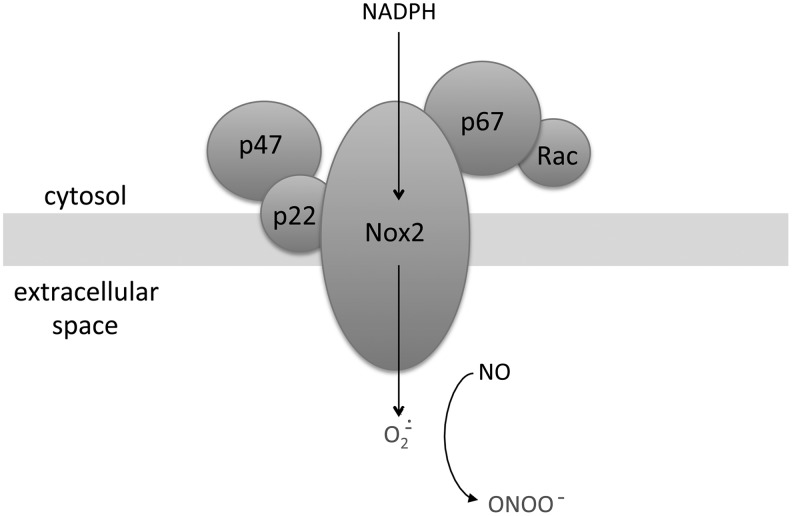
Significance: Abnormal lung development in the perinatal period can result in severe neonatal complications, including persistent pulmonary hypertension (PH) of the newborn and bronchopulmonary dysplasia. Reactive oxygen species (ROS) play a substantive role in the development of PH associated with these diseases. ROS impair the normal pulmonary artery (PA) relaxation in response to vasodilators, and ROS are also implicated in pulmonary arterial remodeling, both of which can increase the severity of PH.

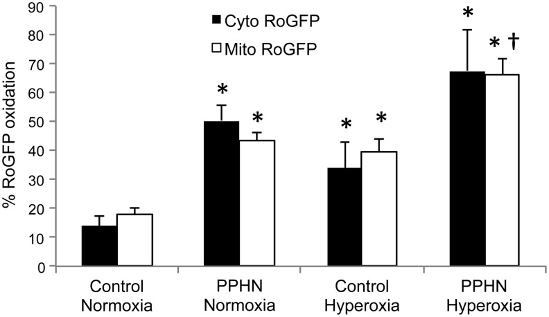
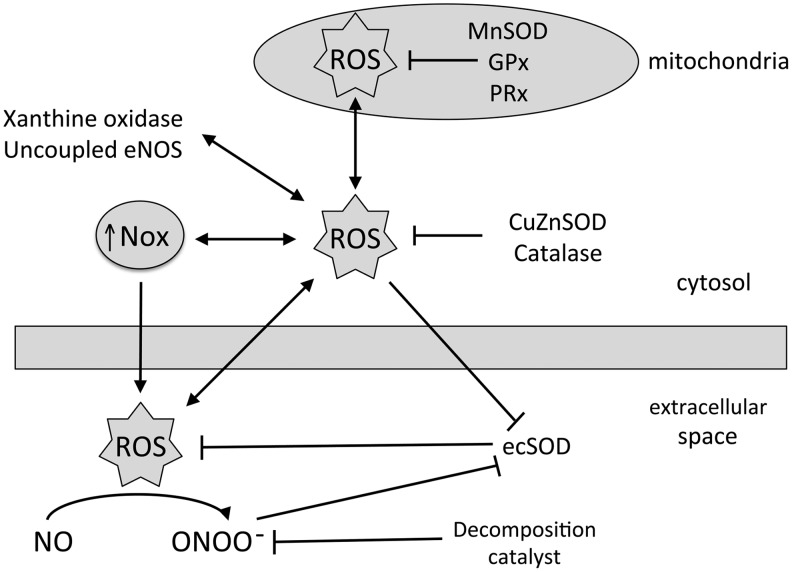
Recent advances: PA ROS levels are elevated when endogenous ROS-generating enzymes are activated and/or when endogenous ROS scavengers are inactivated. Animal models have provided valuable insights into ROS generators and scavengers that are dysregulated in different forms of neonatal PH, thus identifying potential therapeutic targets.

Critical issues: General antioxidant therapy has proved ineffective in reversing PH, suggesting that it is necessary to target specific signaling pathways for successful therapy.

Future directions: Development of novel selective pharmacologic inhibitors along with nonantioxidant therapies may improve the treatment outcomes of patients with PH, while further investigation of the underlying mechanisms may enable earlier detection of the disease.


Figures

<b>FIG. 1.</b>
FIG. 1.
Representative histology of pulmonary vessels from infants who died with severe pulmonary hypertension (PH). (A) Small pulmonary vessel from an infant with asphyxia and pulmonary hypertension of the newborn (PPHN), demonstrating dramatic smooth muscle cell thickening around pulmonary arteries (arrow). (B) Lung photomicrograph with elastin staining from an infant with BPD-associated PH and organizing pneumonia. A thickened medial layer, double elastic lamina, and modest proliferation of the adventitia are noted (arrow). Both examples indicate a lack of the intimal proliferation that characterizes adult PH.
<b>FIG. 2.</b>
FIG. 2.
Mechanisms of pulmonary artery vasodilation and vasoconstriction. Endothelial nitric oxide synthase (eNOS) generates the vasodilator nitric oxide (NO) from L-arginine in endothelial cells (ECs), which activates soluble guanylate cyclase (sGC) in adjacent smooth muscle cells. Active sGC converts guanosine triphosphate (GTP) to cyclic guanosine monophosphate (cGMP), which triggers vasodilation via an enzyme phosphorylation cascade. Conversely, phosphodiesterase type 5 (PDE5) hydrolyzed cGMP to the inactive GMP, thus attenuating the cascade. Endothelin (ET-1) released by ECs triggers a cascade leading to vasoconstriction by activating type a receptors (ETA) on smooth muscle cells.
<b>FIG. 3.</b>
FIG. 3.
Vascular remodeling is associated with increased ROS in PPHN pulmonary arteries. Frozen sections from control and PPHN lamb lungs were incubated with the H2O2-sensitive dye 2′, 7′-dichlorodihydrofluorescein diacetate [adapted from (187)], with the superoxide-sensitive dye dyhydroethidium [adapted from (21)], or fixed and incubated with an antibody to 3-nitrotyrosine residues (3-NT) [adapted from (103)], and visualized by fluorescence microscopy.
<b>FIG. 4.</b>
FIG. 4.
Elevated levels of reactive oxygen species (ROS) induces vasoconstriction and pulmonary vascular remodeling in PPHN via multiple mechanisms. In ECs, ROS inhibit eNOS activity, resulting in decreased levels of bioavailable NO. In smooth muscle cells, ROS inactivate sGC and activate PDE5, resulting in decreased levels of cGMP.
<b>FIG. 5.</b>
FIG. 5.
Diagram illustrating subunit organization of NADPH oxidase 2 (Nox2) at the plasma membrane. Nox2 and p22 are present within the membrane, while p47, p67 and Rac are located in the cytosol. The transfer of electrons from NADPH in the cytosol generates superoxide in the extracellular space. Superoxide decreases bioavailable NO in the rapid reaction and generates the vasoconstrictor peroxynitrite (ONOO).
<b>FIG. 6.</b>
FIG. 6.
Hyperoxia and PPHN increase oxidant stress in the cytosol and mitochondrial matrix of PASMC. PASMC were isolated from control or PPHN lambs and infected with an adenovirus expressing the ROS-sensitive protein RoGFP in the cytosol (Cyto RoGFP) or in the mitochondrial matrix (Mito RoGFP). Cells were exposed to 21% (normoxia) or 95% O2 (hyperoxia) for 24 h and RoGFP oxidation determined by flow cytometry. *p<0.05 versus control normoxia; p<0.05 versus PPHN normoxia. Adapted from (55, 189).
<b>FIG. 7.</b>
FIG. 7.
Diagram depicting the interactions between cellular ROS generators and scavengers. In mitochondria, ROS levels are regulated by enzymes, including manganese superoxide dismutase (MnSOD), glutathione peroxidase (GPx), and peroxyredoxin (PRx). In the cytosol, NADPH oxidases (Nox), xanthine oxidase, and uncoupled endothelial nitric oxide synthase (eNOS) generate ROS, while copper/zinc superoxide dismutase (CuZnSOD) and catalase scavenge ROS. Nox also contribute to ROS in the extracellular space, while extracellular superoxide dismutase (ecSOD) scavenges extracellular superoxide. Increased extracellular superoxide decreases bioavailable NO in the formation of peroxynitrite (ONOO), and this vasoconstrictor is removed in the presence of a decomposition catalyst.

References

    1. Abman SH. Impaired vascular endothelial growth factor signaling in the pathogenesis of neonatal pulmonary vascular disease. Adv Exp Med Biol 661: 323–335, 2010 - PubMed
    1. Abman SH, Shanley PF, and Accurso FJ. Failure of postnatal adaptation of the pulmonary circulation after chronic intrauterine pulmonary hypertension in fetal lambs. J Clin Invest 83: 1849–1858, 1989 - PMC - PubMed
    1. Afolayan AJ, Eis A, Teng RJ, Bakhutashvili I, Kaul S, Davis JM, and Konduri GG. Decreases in manganese superoxide dismutase expression and activity contribute to oxidative stress in persistent pulmonary hypertension of the newborn. Am J Physiol Lung Cell Mol Physiol 303: L870–L879, 2012 - PMC - PubMed
    1. Afshar S, Gibson LL, Yuhanna IS, Sherman TS, Kerecman JD, Grubb PH, Yoder BA, McCurnin DC, and Shaul PW. Pulmonary NO synthase expression is attenuated in a fetal baboon model of chronic lung disease. Am J Physiol Lung Cell Mol Physiol 284: L749–L758, 2003 - PubMed
    1. Ahmed MN, Suliman HB, Folz RJ, Nozik-Grayck E, Golson ML, Mason SN, and Auten RL. Extracellular superoxide dismutase protects lung development in hyperoxia-exposed newborn mice. Am J Respir Crit Care Med 167: 400–405, 2003 - PubMed

Substances

LinkOut - more resources