

doi: 10.1186/1749-8104-8-23.
Brain on the stage - spotlight on nervous system development in zebrafish: EMBO practical course, KIT, Sept. 2013
Affiliations
- PMID: 24350623
- PMCID: PMC3878791
- DOI: 10.1186/1749-8104-8-23
Item in Clipboard
Brain on the stage - spotlight on nervous system development in zebrafish: EMBO practical course, KIT, Sept. 2013
Neural Dev.
.
Abstract
During the EMBO course 'Imaging of Neural Development in Zebrafish', held on September 9-15th 2013, researchers from different backgrounds shared their latest results, ideas and practical expertise on zebrafish as a model to address open questions regarding nervous system development.
Figures
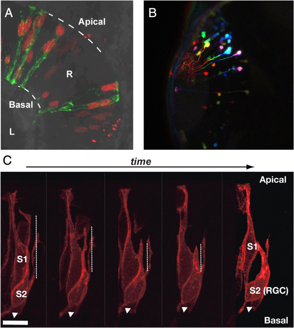
Mosaic labelling techniques in the zebrafish embryo. (A, C) Visualisation of individual transplanted retinal cells from donor embryos labelled with either H2B-RFP and membrane tagged-GFP (A) or the Atoh7-gap43RFP transgene (C) at 28 hpf. In (C) frames from a time-lapse series showing apical process retraction from the apical surface (up, see vertical dotted line) and axon extension at the basal surface (white arrow) of the s2 sibling that differentiates into a retinal ganglion cell after asymmetric division. R, Retina; L, lens. Scale bar is 10 μm. (B) Few small cell clones labelled by electroporation of CMV:mCitrine. Acquired images were pseudo-coloured afterwards according to z-level to visualize individual cell clones in the brain at 32 hpf. In (A, C), the retina is oriented dorsal up. (B) Dorsal view of the midbrain. The data presented in this figure were acquired by the course participants with the help of Wiebke Sassen (Braunschweig), Shahad Albadri and Anne-Laure Duchemin (Heidelberg).
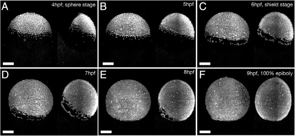
Imaging of zebrafish gastrulation by digital light sheet microscopy. Maximum-intensity projections of H2B-GFP labelled wild type zebrafish embryo at the indicated times and subsequent developmental stages from A to F. The data presented in this figure were acquired by the course participants with the help of Andrey Kobitski, KIT.
Similar articles
-
Making sense of zebrafish neural development in the Minervois.Neural Dev. 2007 Aug 8;2:15. doi: 10.1186/1749-8104-2-15. Neural Dev. 2007. PMID: 17686145 Free PMC article.
-
Slitrk gene duplication and expression in the developing zebrafish nervous system.Dev Dyn. 2014 Feb;243(2):339-49. doi: 10.1002/dvdy.24076. Epub 2013 Nov 21. Dev Dyn. 2014. PMID: 24123428
-
Neurogenesis in the visual system of embryonic and adult zebrafish (Danio rerio). off.Vis Neurosci. 1999 May-Jun;16(3):417-24. doi: 10.1017/s095252389916303x. Vis Neurosci. 1999. PMID: 10349963
-
Zebrafish neural induction and patterning.Dev Dyn. 2000 Oct;219(2):155-68. doi: 10.1002/1097-0177(2000)9999:9999<::aid-dvdy1052>3.3.co;2-b. Dev Dyn. 2000. PMID: 11002336 Review.
-
Somitogenesis.Results Probl Cell Differ. 2002;40:271-97. doi: 10.1007/978-3-540-46041-1_14. Results Probl Cell Differ. 2002. PMID: 12353481 Review. No abstract available.
Cited by
-
Expression of a Barhl1a reporter in subsets of retinal ganglion cells and commissural neurons of the developing zebrafish brain.Sci Rep. 2020 Jun 1;10(1):8814. doi: 10.1038/s41598-020-65435-w. Sci Rep. 2020. PMID: 32483163 Free PMC article.
-
In Vivo Imaging of Transgenic Gene Expression in Individual Retinal Progenitors in Chimeric Zebrafish Embryos to Study Cell Nonautonomous Influences.J Vis Exp. 2017 Mar 22;(121):55490. doi: 10.3791/55490. J Vis Exp. 2017. PMID: 28362422 Free PMC article.
References
Publication types
MeSH terms
LinkOut - more resources
Full Text Sources
Other Literature Sources
