. 2013 Dec 18:1:83.
doi: 10.1186/2051-5960-1-83.

A central role for dityrosine crosslinking of Amyloid-β in Alzheimer's disease

Affiliations

A central role for dityrosine crosslinking of Amyloid-β in Alzheimer's disease

Youssra K Al-Hilaly et al. Acta Neuropathol Commun. .

Abstract

Background: Alzheimer's disease (AD) is characterized by the deposition of insoluble amyloid plaques in the neuropil composed of highly stable, self-assembled Amyloid-beta (Aβ) fibrils. Copper has been implicated to play a role in Alzheimer's disease. Dimers of Aβ have been isolated from AD brain and have been shown to be neurotoxic.

Results: We have investigated the formation of dityrosine cross-links in Aβ42 formed by covalent ortho-ortho coupling of two tyrosine residues under conditions of oxidative stress with elevated copper and shown that dityrosine can be formed in vitro in Aβ oligomers and fibrils and that these links further stabilize the fibrils. Dityrosine crosslinking was present in internalized Aβ in cell cultures treated with oligomeric Aβ42 using a specific antibody for dityrosine by immunogold labeling transmission electron microscopy. Results also revealed the prevalence of dityrosine crosslinks in amyloid plaques in brain tissue and in cerebrospinal fluid from AD patients.

Conclusions: Aβ dimers may be stabilized by dityrosine crosslinking. These results indicate that dityrosine cross-links may play an important role in the pathogenesis of Alzheimer's disease and can be generated by reactive oxygen species catalyzed by Cu2+ ions. The observation of increased Aβ and dityrosine in CSF from AD patients suggests that this could be used as a potential biomarker of oxidative stress in AD.


Figures

Figure 1
Figure 1
Formation of dityrosine crosslinks in Aβ42 fibrils. Preformed Aβ42 fibrils (20 μM) were incubated in the presence of Cu2+/H2O2 for 72 hours and the appearance of dityrosine detected using fluorescence (a). b) The dityrosine content was confirmed using LC-ESIMS/MS and this shown with relative abundance on y-axis. i) LC-ESIMS/MS from authentic synthetic dityrosine, ii) hydrolysate from oxidized preformed Aβ42 fibrils, iii) hydrolysate from Aβ42 fibrils formed under oxidation conditions for three days (the fibrils were obtained from incubation of soluble Aβ42 with Cu2+/H2O2 in water at 37°C and agitation) (c, d) Electron micrographs showing the morphology of fibrils prior to oxidation (c) and following 24 hour oxidation (d) (diameters approximately 100–150 Å).
Figure 2
Figure 2
Monitoring dityrosine and tyrosine fluorescence during Aβ42 assembly. Freshly prepared Aβ42 (20 μM) was incubated in the presence of Cu2+/H2O2 and monitored by fluorescence over three days (a) and compared to Aβ42 alone (b). Dityrosine fluorescence was monitored (Ex. 320 nm, Em. 410–420 nm) and a) oxidized Aβ42 shows a strong signal at 24 hours incubation compared to no signal in b) control Aβ42 sample. The development of the dityrosine signal was monitored at earlier time points (c) using the addition of EDTA and the spectrum shows a signal at 420 nm following only 10 mins incubation. Tyrosine fluorescence (Ex. 280 nm, Em. 305 nm) was used to follow assembly with time (d). The intensity at 305 nm is plotted against time. Both conditions show a decrease in fluorescence signal over time for tyrosine, but oxidized Aβ42 shows a significant reduction after 24 hours, compared to a slow reduction in tyrosine fluorescence that accompanies assembly for control Aβ42.
Figure 3
Figure 3
Thioflavine T fluorescence and SDS PAGE of oxidized and control Aβ42. a) ThT fluorescence spectra following fibril formation from Aβ42 (20 μM) in the presence or absence of Cu2+ and H2O2 for 72 hours. The spectra for oxidized and control samples show an increased intensity over the incubation time and show increased ThT signal for non-oxidized compared to oxidized samples. b) SDS PAGE showing separation of Aβ42. Oxidized Aβ42 (left column) runs as monomer and dimer (approx. 9 kDa, black arrow), whilst non oxidized, control Aβ42 (right column) shows bands corresponding to monomer and trimer as previously observed [44]. The trimer is thought to be induced by SDS [44]. Densitometry confirms that monomer is the strongest band following by dimer for oxidized but not control fibrils. This reveals that the dimer is enriched under oxidation conditions.
Figure 4
Figure 4
Transmission electron microscopy images of freshly formed Aβ42 fibrils. 20 μM Aβ42 (pH 7.4) was incubated in the presence of Cu2+/H2O2(a, b, c) and compared to 20 μM Aβ42 in phosphate buffer alone (pH 7.4) (d, e, f). Assembly was monitored by electron microscopy after incubation for (a and d) zero hours, (b and e) 24 hours and (c and f) 48 hours. Both oxidized and control Aβ42 samples show oligomeric species at zero hour (a, d) and fibrils following 48 hour incubation (c, g). However, at 24 hours (b and f) fibrils were observed in non-oxidized conditions (f), but no fibrils were observed in oxidised conditions (b). The presence of dityrosine was detected using immunogold labeling using a dityrosine specific antibody that labeled oxidized fibrils (g) but not control fibrils (h).
Figure 5
Figure 5
The stability of crosslinked fibrils. a) Oxidized and non-oxidized Aβ42 fibrils were examined using dityrosine fluorescence after prolonged incubation at −80°C showing a strong intensity signal at 420 nm for oxidized but not non-oxidized fibrils. b) Formic acid was used to dissolve the fibrils and the concentration of Aβ42 in solution was compared before and after formic acid dissolution for oxidized and non-oxidized fibrils. c) Electron micrographs of oxidized fibrils before and after formic acid treatment showing that the dityrosine crosslinked fibrils are resistant to formic acid. d) Electron micrographs of non-oxidized fibrils before and after formic acid treatment showing the fibrils are susceptible to damage by formic acid. Scale bars represent 0.2 μm.
Figure 6
Figure 6
Immunogold labeling TEM showing neuroblastoma cells treated with 10 μM oligomeric Aβ. a) The images reveal dityrosine (10 nm) and Aβ42 (5 nm) labeling within the lysosomes of treated cells. b) low level dityrosine labeling was observed within lysosomes in vehicle treated, control cells, but no Aβ labeling was observed. c) Cells were observed containing fibrillar Aβ labeled for both Aβ (5 nm) and dityrosine (10 nm). White arrows are used to highlight the fibrillar material. Inserts show magnified images for further clarity.
Figure 7
Figure 7
Electron micrographs of sections of Aβ42 treated neuroblastoma cells showing immunogold labeling of Aβ (5 nm) and dityrosine (10 nm). The images reveal colabeled fibrils outside and also inside the cells and highlight internalization at the plasma membrane (top left panel). The figure shows magnified images in the bottom two panels for extra clarity.
Figure 8
Figure 8
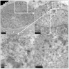
Immunogold labeling TEM within amyloid plaques from AD brains. a) Shows double labeling using the dityrosine antibody and anti-Aβ antibody on an AD brain section and reveals very dense labeling of the amyloid plaques. This is compared to a serial section b) Showing single labelling using an irrelevant antibody against hair cell antigen (HCA) to show the specificity of the dityrosine labeling on plaques. This image shows virtually no gold labeling. c) Shows single labeling using the dityrosine antibody to reveal very clear labeling at increasing magnification (ci, cii and ciii) showing that the dityrosine labels fibrils in the plaques. d) Double labeling with anti-Aβ (5 nm) and anti-dityrosine (10 nm) confirms colocalisation of dityrosine with Aβ within amyloid plaques at increasing magnifications (di and dii). e) Shows the 10 nm labeling for dityrosine highlighted in red and 5 nm labeling for Aβ in blue. Inserts show magnified images for further clarity. f) and g) show additional examples of co-immunogold labeling in brain from additional patients.
Figure 9
Figure 9
Immunogold labeling TEM/negatively stain of cerebrospinal fluid from AD brains and age matched controls. a) A higher density of dityrosine (labeled with 15 nm) and Aβ (labeled with 5 nm) labeling was observed for CSF from an AD patient (top images) compared to b) age matched control. In CSF of AD patients, we can identify two different areas of labeling, low-density co-localization (☐), and high density co-localization (◯) of dityrosine and Aβ. We observe significant labeling levels for AD CSF compared to virtually no labeling in CSF from control patients. Inserts show magnified images for further clarity.
Figure 10
Figure 10
Schematic summarizing the mechanism of dityrosine formation in Aβ in complex with copper including the generation of ROS and summarizing the results showing the physiological importance of dityrosine crosslinked Aβ in Alzheimer’s disease.

References

    1. Selkoe DJ. The molecular pathology of Alzheimer’s disease. Neuron. 1991;1:487–498. doi: 10.1016/0896-6273(91)90052-2. - DOI - PubMed
    1. Rambaran RN, Serpell LC. Amyloid fibrils: abnormal protein assembly. Prion. 2008;1:112–117. doi: 10.4161/pri.2.3.7488. - DOI - PMC - PubMed
    1. Hardy J, Selkoe DJ. The amyloid hypothesis of Alzheimer’s disease: progress and problems on the road to therapeutics. Science. 2002;1:353–356. doi: 10.1126/science.1072994. - DOI - PubMed
    1. Luhrs T, Ritter C, Adrian M, Riek-Loher D, Bohrmann B, Dobeli H, Schubert D, Riek R. 3D structure of Alzheimer’s amyloid-beta(1–42) fibrils. Proc Natl Acad Sci USA. 2005;1:17342–17347. doi: 10.1073/pnas.0506723102. - DOI - PMC - PubMed
    1. Hensley K, Maidt ML, Yu Z, Sang H, Markesbery WR, Floyd RA. Electrochemical analysis of protein nitrotyrosine and dityrosine in the Alzheimer brain indicates region-specific accumulation. J Neurosci. 1998;1:8126–8132. - PMC - PubMed

Publication types