Review
. 2013 Dec 17;5(12):2533-71.
doi: 10.3390/toxins5122533.

Secreted phospholipases A2 of snake venoms: effects on the peripheral neuromuscular system with comments on the role of phospholipases A2 in disorders of the CNS and their uses in industry

Affiliations
Review

Secreted phospholipases A2 of snake venoms: effects on the peripheral neuromuscular system with comments on the role of phospholipases A2 in disorders of the CNS and their uses in industry

John B Harris et al. Toxins (Basel). .

Abstract

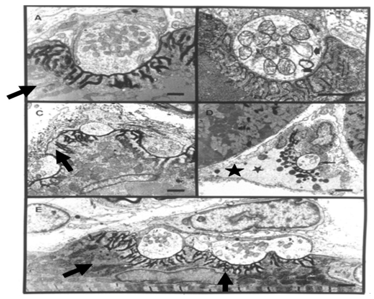
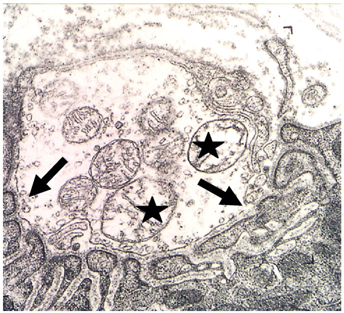
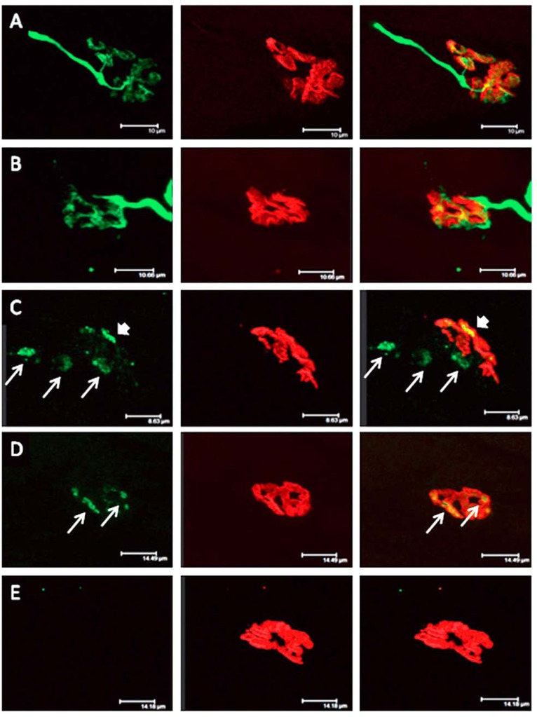
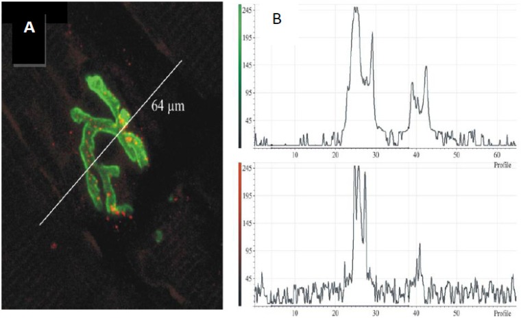
Neuro- and myotoxicological signs and symptoms are significant clinical features of envenoming snakebites in many parts of the world. The toxins primarily responsible for the neuro and myotoxicity fall into one of two categories--those that bind to and block the post-synaptic acetylcholine receptors (AChR) at the neuromuscular junction and neurotoxic phospholipases A2 (PLAs) that bind to and hydrolyse membrane phospholipids of the motor nerve terminal (and, in most cases, the plasma membrane of skeletal muscle) to cause degeneration of the nerve terminal and skeletal muscle. This review provides an introduction to the biochemical properties of secreted sPLA2s in the venoms of many dangerous snakes and a detailed discussion of their role in the initiation of the neurologically important consequences of snakebite. The rationale behind the experimental studies on the pharmacology and toxicology of the venoms and isolated PLAs in the venoms is discussed, with particular reference to the way these studies allow one to understand the biological basis of the clinical syndrome. The review also introduces the involvement of PLAs in inflammatory and degenerative disorders of the central nervous system (CNS) and their commercial use in the food industry. It concludes with an introduction to the problems associated with the use of antivenoms in the treatment of neuro-myotoxic snakebite and the search for alternative treatments.


Figures

Figure 1
Figure 1
Glycerophospholipid structure and the site of action of phospholipases. The phospholipid molecule consists of a glycerol-3-phosphate (blue) esterified at positions sn-1 and sn-2 to non-polar fatty acids. Its phosphoryl group is esterified to a polar head group (x). Phospholipases A1 cleave the ester bond at the sn-1 position. Phospholipases A2 cleave the ester bond at the sn-2 position. Phospholipases B cleave the ester bonds at both sn-1 and sn-2 positions. Phospholipases C cleave the glycerophosphate bond and phospholipases D remove the polar head group. From The AOCS Lipid Library (http://lipidlibrary.aocs.org/animbio/phospholipases/index.htm).
Figure 2
Figure 2
Snakes frequently involved in major neuro-myotoxic envenoming in humans. (A) the Australian Tiger snake, Notechis scutatus and (B) the South American rattlesnake, Crotalus durissus terrificus, both cause neurotoxicity and rhabdomyolysis; (C) the Taiwanese Multi-banded krait, Bungarus multicinctus causes severe neurotoxicity but no myotoxicity; (D) the Beaked sea snake, Enhydrina schistosa, causes severe myotoxicity but rarely neurotoxicity in human subjects.
Figure 3
Figure 3
(A). A group 1A phospholipase A2 with phospholipid substrate modeled in the active site. The active site residues His-48 and Asp-99 and the bound Ca2+ is shown in purple. Ca2+ is bound by Asp-49 as well as the carbonyl oxygens of Tyr-28, Gly-30 and Gly-32. Aromatic residues are shown in white; (B). Model of the lipid binding of the group 1A PLA2 is shown with residues on the interfacial binding surface Tyr-3, Trp-19, Trp-61 and Phe-64 shown in stick form. From Burke and Dennis 2008 [1].
Figure 4
Figure 4
Victim of an envenoming bite by an unidentified snake on admission at a tertiary referral hospital in Chittagong, Bangladesh. Note multiple tight ligatures applied to the arm.
Figure 5
Figure 5
Incisions applied to the hand and lower leg respectively in two victims of envenoming bites by unidentified snakes in Chittagong, Bangladesh.
Figure 6
Figure 6
Localised necrosis following an envenoming bite to the foot by the viperid Fer de Lance, (Bothrops asper).
Figure 7
Figure 7
(A) Young boy with severe neurotoxic signs following a bite by a cobra (species unknown) in Bangladesh; (B) Full recovery 24 h later following treatment with antivenom.
Figure 8
Figure 8
Wrist drop and foot drop, respectively, many months after the apparently successful treatment of victims of neurotoxic snake bites in Chittagong, Bangladesh.
Figure 9
Figure 9
Severe neurotoxicity and rhabdomyolysis (note the black urine) following an envenoming bite by a greater black krait, Bungarus niger in Bangladesh. The patient did not recover.
Figure 10
Figure 10
Ptosis and rhabdomyolysis (note the black urine) following an envenoming bite by South American rattlesnake (Crotalus durissus) in Brazil.
Figure 11
Figure 11
TEM Images of motor nerve terminal boutons on muscle fibres of the rat 12–24 h after the inoculation of notexin, a PLA2 toxin from the venom of the Australian tiger snake, Notechis scutatus. (A) Control bouton on a muscle fibre not exposed to any toxin. Note the folds of the postsynaptic membrane (Arrows); (BE) Note the widespread loss of synaptic vesicles from the boutons and the swollen mitochondria (small arrows). Note also the well preserved junction folds of the neuromuscular junctions (large arrows in C). Combined damage to both bouton and muscle fibre is shown in D: a star marks the collapsed muscle fibre but note the preservation of the junctional folds at the neuromuscular junction.
Figure 12
Figure 12
A terminal bouton in advanced stages of degeneration. Note the lesions in the plasma membrane (arrows) and the damaged mitochondria (stars).
Figure 13
Figure 13
Longitudinal sections of rat soleus muscles 24 h after the inoculation, in vivo, of the venom of the Greater black krait, Bungarus niger. Sections were labelled with TRITC-conjugated α-Bungarotoxin to label junctional ACh receptors (red) and FITC conjugated Ab to neurofilament protein to label motor axons (green). (A) control image; (BE) Progressive breakdown of the terminal innervation at the neuromuscular junction. Note the preservation of the junctional ACh receptors (From Faiz et al. 2010 [68]). Reproduced with permission from Publisher.
Figure 14
Figure 14
(A) Longitudinal section of murine muscle labelled with ammodytoxin A, an sPLA2 from the venom of the long-nosed viper, Vipera ammodytes, conjugated with Alexa546 (red) and counter-labelled with FITC-conjugated α-Bungarotoxin to label junctional ACh receptors (green) (B) a laser scan of red and green channels to demonstrate localisation of sPLA2 to the neuromuscular junction. (From Logonder et al. 2008 [85]). Reproduced with permission from the Publisher.
Figure 15
Figure 15
The density of synaptic vesicles in terminal boutons of rat neuromuscular junctions. Vesicle density was unchanged in muscles incubated in vitro with either botulinum toxin C or conotoxin ω-MVIIC. Incubation with β-bungarotoxin, an SPLA2 toxin from the venom of Bungarus multicinctus, caused a significant fall in vesicle density. The fall was largely or completely prevented in muscles pre-treated with either botulinum toxin C or conotoxin ω-MVIIC before exposure to β-bungarotoxin. (From Prasarnpun et al. 2004 [76]). Reproduced with permission from the Publisher.
Figure 16
Figure 16
TEM images of terminal boutons on murine muscle fibres previously exposed to a gold-labelled sPLA2 from the venom of the horned viper (Vipera ammodytes ammodytes). The control bouton (A) is not decorated; Bouton B shows particles within the synaptic cleft and folds. Bouton C shows particles in the synaptic cleft and the bouton itself. Enlarged images (DI) show particles associated with vesicle–like structures within the bouton or with mitochondria (open arrows). The association between label and vesicle-like structures suggest that uptake might occur during the recycling of synaptic vesicles and endocytosis. (Modified from Logonder et al. 2009 [94]). Reproduced with permission from the Publisher.
Figure 17
Figure 17
A cluster of six individual end-plates (labelled with FITC-conjugated α-bungarotoxin) innervated by the clustered intramuscular branching of a single motor axon (labelled with TRITC-conjugated anti-neurofilament Ab.
Figure 18
Figure 18
(AC) Transverse sections of soleus muscles stained with haematoxylin and eosin (H&E). (A) control; (B,C) Three and 24 h respectively after exposure, in vivo, to notexin, an sPLA2 from the venom of the Australian tiger snake, Notechis scutatus. Note the early inflammatory response and the later degeneration of the muscle fibres; (D) Longitudinal section at 24 h stained with procion yellow. This dye is excluded from cells with an intact plasma membrane. Note that it has entered the muscle fibres and stained the congealed, hyper-contracted myofilaments.
Figure 19
Figure 19
TEM of a longitudinal section of a rat soleus muscle fibre labelled with a gold-conjugated Ab against notexin, an sPLA2 from the venom of the Australian tiger snake, Notechis scutatus, three hrs after exposure in vivo to the toxin. Arrows indicate individual silver-enhanced gold particles. (From Dixon and Harris 1996 [109]). Reproduced with permission from the Publisher.
Figure 20
Figure 20
The relative rates of loss of desmin and myosin from muscles at various times after the inoculation of the venom of Notechis scutatus. (From Harris et al. 2003 [114]). Reproduced with permission from the Publisher.
Figure 21
Figure 21
(AC) Transverse sections of soleus muscles stained with H&E. A. control. B, C. Four and 28 days respectively after exposure, in vivo, to notexin, an sPLA2 from the venom of the Australian tiger snake, Notechis scutatus. Note the rapid growth of the muscle fibres and the continuing presence of centrally located myonuclei; (D) As above 28 days after exposure in vivo to the venom of the Fer de Lance, Bothrops asper, a viperid snake that causes extensive soft tissue necrosis (see Figure 5). Note the immature appearance of the regenerating muscle fibres and the extensive infiltration of connective tissue.

Similar articles

Cited by

References

    1. Burke J.E., Dennis E.A. Phopholipase A2 structure/function, mechanism and signaling. J. Lipid Res. 2008;50:S237–S242. doi: 10.1194/jlr.R800033-JLR200. - DOI - PMC - PubMed
    1. Dennis E.A., Cao J., Hsu Y.-H., Magrioti V., Kokotos G. Phospholipase A2 enzymes: Physical structure, biological function, disease implication, chemical inhibition and therapeutic intervention. Chem. Rev. 2011;111:6130–6185. doi: 10.1021/cr200085w. - DOI - PMC - PubMed
    1. Leslie C.C. Properties and regulation of cytosolic phospholipase A2. J. Biol. Chem. 1997;272:16709–16712. doi: 10.1074/jbc.272.27.16709. - DOI - PubMed
    1. Herkert M., Shakhman O., Schweins E., Becker C.-M. β-Bungarotoxin is a potent inducer of apoptosis in cultured rat neurons by receptor-mediated internalization. Eur. J. Neurosci. 2001;14:821–828. doi: 10.1046/j.0953-816x.2001.01699.x. - DOI - PubMed
    1. Kalyvas A., David S. Cytosolic phospholipases A2 plays a key role in the pathogenesis of multiple sclerosis-like disease. Neuron. 2004;41:323–335. doi: 10.1016/S0896-6273(04)00003-0. - DOI - PubMed

Publication types

LinkOut - more resources