Review
. 2013 Dec 16;14(12):24438-75.
doi: 10.3390/ijms141224438.

Oxidative stress and neurodegenerative disorders

Affiliations
Review

Oxidative stress and neurodegenerative disorders

Jie Li et al. Int J Mol Sci. .

Abstract

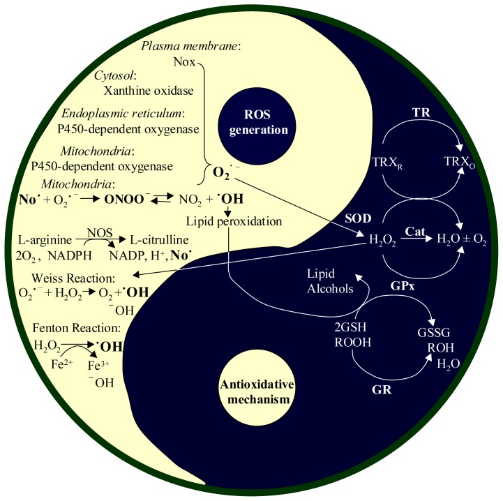
Living cells continually generate reactive oxygen species (ROS) through the respiratory chain during energetic metabolism. ROS at low or moderate concentration can play important physiological roles. However, an excessive amount of ROS under oxidative stress would be extremely deleterious. The central nervous system (CNS) is particularly vulnerable to oxidative stress due to its high oxygen consumption, weakly antioxidative systems and the terminal-differentiation characteristic of neurons. Thus, oxidative stress elicits various neurodegenerative diseases. In addition, chemotherapy could result in severe side effects on the CNS and peripheral nervous system (PNS) of cancer patients, and a growing body of evidence demonstrates the involvement of ROS in drug-induced neurotoxicities as well. Therefore, development of antioxidants as neuroprotective drugs is a potentially beneficial strategy for clinical therapy. In this review, we summarize the source, balance maintenance and physiologic functions of ROS, oxidative stress and its toxic mechanisms underlying a number of neurodegenerative diseases, and the possible involvement of ROS in chemotherapy-induced toxicity to the CNS and PNS. We ultimately assess the value for antioxidants as neuroprotective drugs and provide our comments on the unmet needs.


Figures

Figure 1.
Figure 1.
ROS generations and antioxidant systems in cells. Reactive oxygen species (ROS) can be generated from various sites in a cell. The cell protects its own damage from excessive ROS by suppression of ROS levels by two redox buffers (glutathione (GSH) and thioredoxin (TRX)) and antioxidant enzymes, including superoxide dismutase (SOD), catalase (CAT), thioredoxin reductase (TR), and glutathione peroxidase (GPx). In the cell, superoxide dismutase (SOD) first converts O2•− into H2O2, and then catalase (Cat) enzymatically converts H2O2 is into H2O and O2. In the GSH buffer system, GPx converts H2O2 into H2O and O2 when it converts GSH into its oxidized disulfide form (GSSG). GSSH is then reduced by glutathione reductase (GR) to regenerate GSH for reuse. In the TRX buffering system, the TRX in reduced status (TRXR) is oxidized into oxidized thioredox (TRXO) during the degradation of H2O2 and then reduced by TR. Besides ROS, No is also enzymatically generated by nitric oxide synthases (NOS), and is further reacted with O2•− to produce ONOO.
Figure 2.
Figure 2.
Contribution of ROS to the process of learning and memory. Long-term potentiation (LTP) is considered one of the major cellular mechanisms for learning and memory. It is positively regulated by activation of glutamate receptors, including N-methyl-d-aspartate receptor (NMDAr), α-amino-3-hydroxy-5-methyl-4-isoxazolepropionic acid receptor (AMPAr) and metabotropic glutamate receptor (mGluR). The activation of these receptors results in calcium influx, which then activates different kinases in the cascade to facilitate LTP formation. In contrast, LTP is negatively regulated by phosphatases, including protein phosphatase 2A and 2B (PP2A and PP2B). Activation of glutamate receptors result in O2•− accumulation, possibly through the conversion of NADPH oxidase (Nox).
Figure 3.
Figure 3.
The causal relationship between ROS and misfolded proteins underlying neurodegenerative diseases. A common feature in neurodegenerative diseases is the existence of hallmark protein(s) for each, such as Tau and beta-amyloid (Aβ) for Alzheimer’s disease (AD), alpha-synuclein (αSyn) for Parkinson’s disease, mutant huntingtin protein (mHtt) for Huntington’s disease, and TAR DNA binding protein (TDP-43) for Amyotrophic lateral sclerosis. ROS mediate neurotoxicity in each of these diseases through modifying the hallmark protein by oxidation. In AD, ROS could also activate c-Jun N-terminal kinases (JNK) and p38, and deactivate protein phosphatase 2A (PP2A). JNK and p38 promote the expression of Tau, which is inhibited by PP2A. The activation of JNK and p38 further stimulate AβPP cleaving enzyme 1 (BACE1), causing Aβ1-42 accumulation, which leads to activation of NADPH oxidase (Nox) to produce additional O2•−, and results in Ca2+ influx to elicit excitatory neurotoxicity. In Huntington’s disease, aggregation of mHtt could inhibit peroxiredoxin (Prx), which is an antioxidant.
Figure 4.
Figure 4.
Effect of Antioxidants (AO) on the Neurotoxicity of 5-Fluorouracil (5-FU). (A) Morphological observation. Embryonic brain neurons were cultured for 13 days with concentration reduction of AO in culture medium to 25% and 12.5%, respectively. This process did not elicit cell death by itself (comparison of Non-Insult groups under higher and lower AO conditions). Treating neurons with 25 μM 5-FU did not cause observable neurotoxicity under 25% AO culture condition. However, 5-FU at the same concentration resulted in severe cell death when AO concentration was reduced to 12.5%, even for a shorter time periods. The cells were photographed under a phase-contrast microscope using a 10× objective; and (B) Quantification of cytotoxicity. 3-(4,5-Dimethylthiazol-2-yl)-5-(3-carboxymethoxyphenyl)-2-(4-sulfophenyl)-2H-tetrazolium, inner salt (MTS) was used for the quantitative assay, which directly indicates an enzymatic activity of mitochondria and indirectly reflects the survival cell number. The graph shows that the neurons under reduced AO condition were vulnerable to 5-FU. Data are expressed as mean (n = 6) percentage of Non-Insult groups (taken as 100%). 5-FU dose-dependently reduced MTS reading in reduced AO condition.

References

    1. Turrens J.F. Mitochondrial formation of reactive oxygen species. J. Physiol. 2003;552:335–344. - PMC - PubMed
    1. Coon M.J., Ding X., Pernecky S.J., Vaz A.D.N. Cytochrome P450: Progress and predictions. FASEB J. 1992;6:669–673. - PubMed
    1. Reed J.R., Backes W.L. Formation of P450·P450 complexes and their effect on P450 function. Pharmacol. Ther. 2012;133:299–310. - PMC - PubMed
    1. DeLeo F.R., Quinn M.T. Assembly of the phagocyte NADPH oxidase: Molecular interaction of oxidase proteins. J. Leukoc. Biol. 1996;60:677–691. - PubMed
    1. Finkel T. Redox-dependent signal transduction. FEBS Lett. 2000;476:52–54. - PubMed

LinkOut - more resources