. 2013:4:2965.
doi: 10.1038/ncomms3965.

Structural insights into the role of the Smoothened cysteine-rich domain in Hedgehog signalling

Affiliations

Structural insights into the role of the Smoothened cysteine-rich domain in Hedgehog signalling

Rajashree Rana et al. Nat Commun. 2013.

Abstract

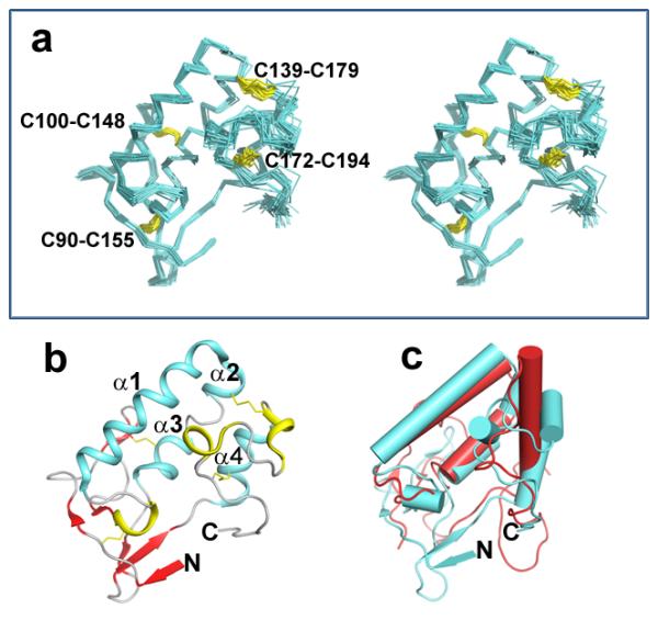
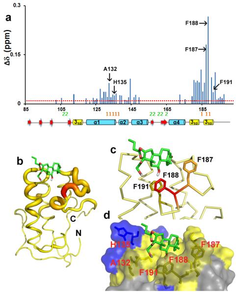
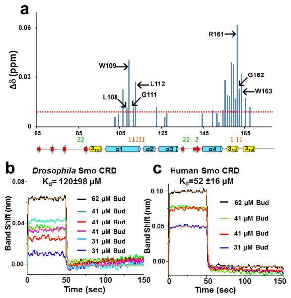
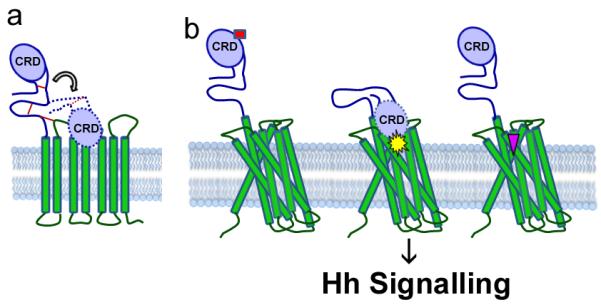
Smoothened (Smo) is a member of the Frizzled (FzD) class of G-protein-coupled receptors (GPCRs), and functions as the key transducer in the Hedgehog (Hh) signalling pathway. Smo has an extracellular cysteine-rich domain (CRD), indispensable for its function and downstream Hh signalling. Despite its essential role, the functional contribution of the CRD to Smo signalling has not been clearly elucidated. However, given that the FzD CRD binds to the endogenous Wnt ligand, it has been proposed that the Smo CRD may bind its own endogenous ligand. Here we present the NMR solution structure of the Drosophila Smo CRD, and describe interactions between the glucocorticoid budesonide (Bud) and the Smo CRDs from both Drosophila and human. Our results highlight a function of the Smo CRD, demonstrating its role in binding to small-molecule modulators.


Figures

Figure 1
Figure 1. Sequence alignment of the Smo CRD
Primary sequence alignment of the Drosophila (D), human (H), mouse (M) and chicken (C) Smo CRD with that of mouse FzD8 CRD and mouse secreted Frizzled Related Protein 3 (sFRP3). The residues conserved in Smo and FzD CRD are shown in red, whereas the residues conserved only in the Smo CRD are shown in blue. The cysteine in FzD not conserved in Smo is underlined and in green. The disulphide bond pattern for the Smo CRD is shown in thick purple lines. The secondary structure elements are shown above and below the primary sequence. The residues highlighted in orange indicate the “site 1” residues of mouse FzD8 that interact with the palmitate modification on the ligand Wnt. The residues highlighted in green indicate the “site 2” on mouse FzD8 that interact with the amino acid sidechains on the opposite side of Wnt. The sequence alignment was generated using ClustalW2.
Figure 2
Figure 2. Identification of cysteines essential for Smo signalling
CRD residues C90, C139, C155, C179 and the Smo ECLD residues C218, C238 and C242 are required for proper Hh reporter gene induction. Mutation of C84, 100,148,172,194,203 to alanine did not compromise the ability of Smo protein to induce the Hh reporter gene activity, and behaved similarly to the wild type protein. Percent activity for each of the experimental assays is shown relative to control. The control level of Hh-induced, ptcΔ136-luciferase activity for control dsRNA was set to 100%. For all conditions, luciferase activity is normalized to pAc-renilla control. Experiments were performed a minimum of two times in duplicate or triplicate and all data pooled. Error bars indicate standard error of the mean (s.e.m.).
Figure 3
Figure 3. Solution structure of the Smo CRD
(a) Stereo view of the backbone atoms (N, Cα, C') of the 20 superimposed structures of Smo CRD with the lowest energy. The disulphides are indicated in yellow and are labelled for clarity. (b) Ribbon diagram of the Smo CRD showing the secondary structure elements. The structure with the lowest energy is used to describe the secondary structure elements. The colour scheme is as follows: Cyan: Alpha helices; Red: β strands; Yellow: 310 helices Grey: random coil. (c) Superimposition of the Smo CRD with FzD CRD. The Smo CRD is represented in cyan and the FzD8 CRD is represented in red. The helices in both proteins are shown as cylinders and the beta-strands as arrowheads. All figures were generated using PyMol.
Figure 4
Figure 4. Sub cellular localization of the cysteines essential for Smo signalling
(a) CRD mutants with compromised signalling activity have altered sub cellular localization. Cl8 cells expressing wild type or the indicated Myc-Smo mutant protein, in the presence of Hh (+) or empty vector control, were examined by indirect immunofluorescence. Wild type Smo translocates to the plasma membrane in response to Hh, whereas Smo CRD cysteine to alanine mutant C90A that was required for maximal Hh reporter gene induction was largely retained in the ER. Smo was detected using anti-Myc (red), Calreticulin-GFP-KDEL marks the ER (green) and DAPI (blue) marks the nucleus. Scale bar: 10uM (b) Sequence alignment of the Drosophila (D) Smo ECLD and ECL1 with the human (H) Smo ECLD and ECL1. The cysteines engaged in disulphide bond formation in human Smo are conserved in Drosophila. The red lines indicate the disulphide bond pattern.
Figure 5
Figure 5. Drosophila Smo CRD binds to the glucocorticoid Bud
(a) CSPs of Smo CRD upon addition of Bud are plotted versus residue numbers. The red line indicates CSP greater than 0.01 ppm. The residues labelled in black form the Bud-binding pocket on Smo CRD as analysed from the HADDOCK docking experiments. The mouse FzD8-Wnt interacting “site 1” and “site 2” residues are shown in orange and green respectively. The corresponding secondary structure elements of the Drosophila Smo CRD are represented below the plot. (b) “Ribbon” representation of the Smo CRD. The backbone thickness of the ribbon is directly proportional to the weighted sum (in ppm) of the 1H and 15N chemical shifts upon binding to the ligand Bud. (c and d) Results of the HADDOCK docking of Bud on Smo CRD. (c) The aromatic side chains of the Bud contacting Smo CRD residues are shown. (d) Surface representation of the residues that interact with Bud are shown in yellow and the positively charged H135 is shown in blue.
Figure 6
Figure 6. Analysis of the binding of Bud to human Smo CRD
(a) CSPs of human Smo CRD upon addition of Bud are plotted versus residue numbers. Dotted red line indicates CSP greater than 0.01 ppm. L108, W109, G111, L112 and R161 are homologous to the mouse Smo residues that interact with 20-OHC. G162 is homologous to Drosophila F187 and W163 is homologous to Drosophila Smo F188 that interacts with Bud. All these residues map to the “site 1” of mouse FzD8-Wnt interaction. “Site 1” and “site 2” residues of mouse FzD8-Wnt interaction are shown in orange and green respectively. The secondary structure elements as in Drosophila Smo CRD structure are shown below the plot. (b) BLI binding assays show that Bud binds to Drosophila Smo CRD. The super streptavidin sensors with biotinylated Drosophila Smo CRD were exposed to three different concentrations of Bud (62, 41, and 31 μM). The processed data were fitted locally with the integrated fitting function by a 1:1 binding model (orange line). The respective Kd values obtained by curve fitting were 89 μM (62 μM Bud, black line), 74 μM (41 μM Bud, green line), 59 μM (41 μM Bud, red line), 318 μM (41 μM Bud, magenta line), 93 μM (31 μM Bud, cyan line), and 85 μM (31 μM Bud, blue line). The average Kd value of Drosophila Smo CRD for Bud was 120 ± 98 μM. (c) BLI binding assays show that Bud binds to human Smo CRD. The experimental and data analysis procedure were same as described above. The respective Kd values obtained by curve fitting were 74 μM (62 μM Bud, black line), 37 μM (41 μM Bud, green line), 44 μM (41 μM Bud, red line) and 54 μM (31 μM Bud, blue line). The average Kd value of human Smo CRD for Bud was 52 ± 16 μM.
Figure 7
Figure 7. A novel model for Smo allosteric regulation
(a) Smo CRD may be flexible. The disulphide bonds stabilizing Smo extracellular linker and extracellular loop are shown in red lines. (b) Smo has more than one binding site. We propose that molecules like Bud bind to the Smo CRD (left, red rectangle) to alter its conformation and attenuate its signalling activity. Cyclopamine and vismodegib are known to bind near the orthosteric binding site located in the cavity of the Smo 7TM domains (right, pink inverted triangle). We speculate that there is the possibility of a class of molecules (middle, yellow star) which would bind to the CRD and cause a conformational change of the Smo extracellular structures to bring it closer to the 7TM domains. This in turn might change the conformation of the trans-membrane domains to regulate signalling.

Similar articles

Cited by

References

    1. Nüsslein-Volhard C, Wieschaus E. Mutations affecting segment number and polarity in Drosophila. Nature. 1980;287:795–801. - PubMed
    1. van den Heuvel M, Ingham PW. smoothened encodes a receptor-like serpentine protein required for hedgehog signalling. Nature. 1996;382:547–551. - PubMed
    1. Alcedo J, Ayzenzon M, VonOhlen T, Noll M, Hooper JE. The Drosophila smoothened gene encodes a seven-pass membrane protein, a putative receptor for the hedgehog signal. Cell. 1996;86:221–232. - PubMed
    1. Ogden SK, et al. G protein G alphai functions immediately downstream of Smoothened in Hedgehog signalling. Nature. 2008;456:967–976. - PMC - PubMed
    1. Ingham PW, McMahon AP. Hedgehog signaling in animal development: paradigms and principles. Genes Dev. 2001;15:3059–3087. - PubMed

Publication types

Associated data