. 2014 Mar;88(5):2530-43.
doi: 10.1128/JVI.03235-13. Epub 2013 Dec 18.

Nucleic acid chaperone activity associated with the arginine-rich domain of human hepatitis B virus core protein

Affiliations

Nucleic acid chaperone activity associated with the arginine-rich domain of human hepatitis B virus core protein

Tien-Hua Chu et al. J Virol. 2014 Mar.

Abstract

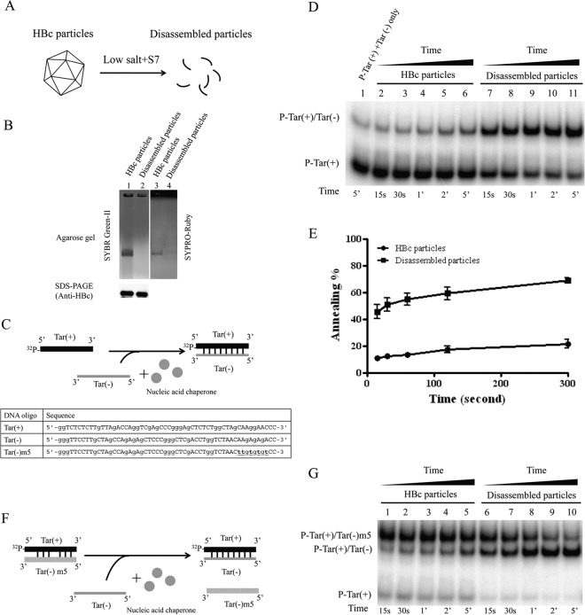
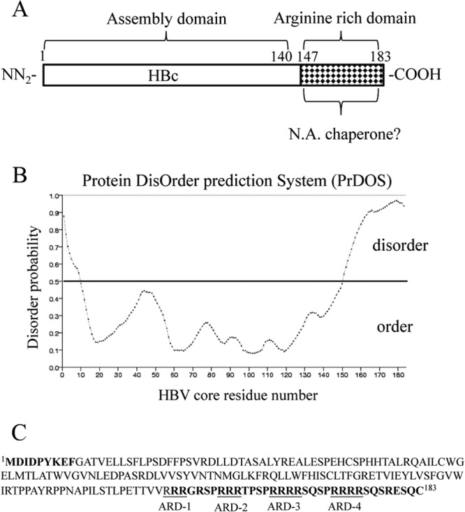
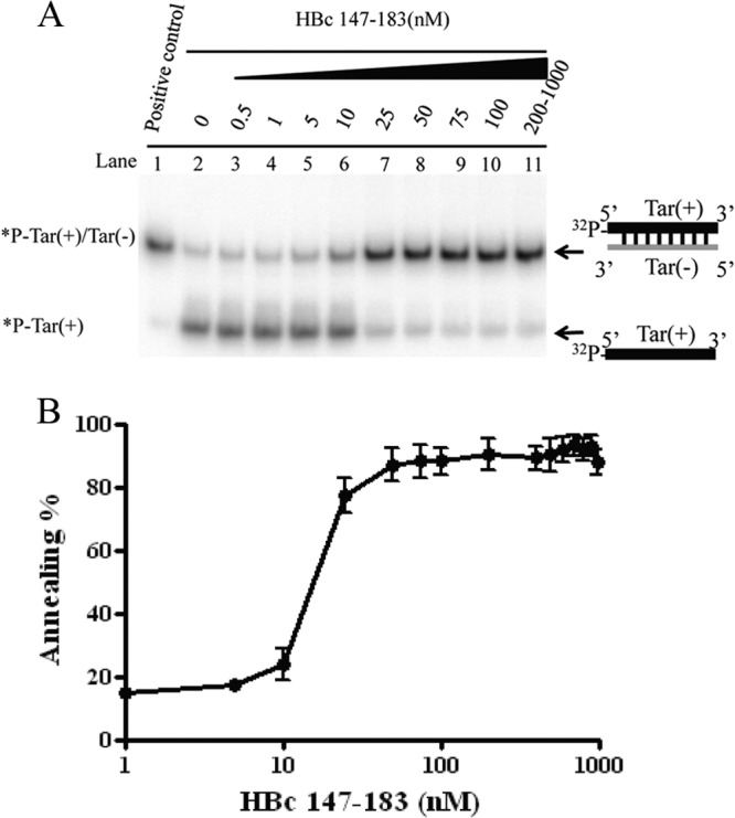
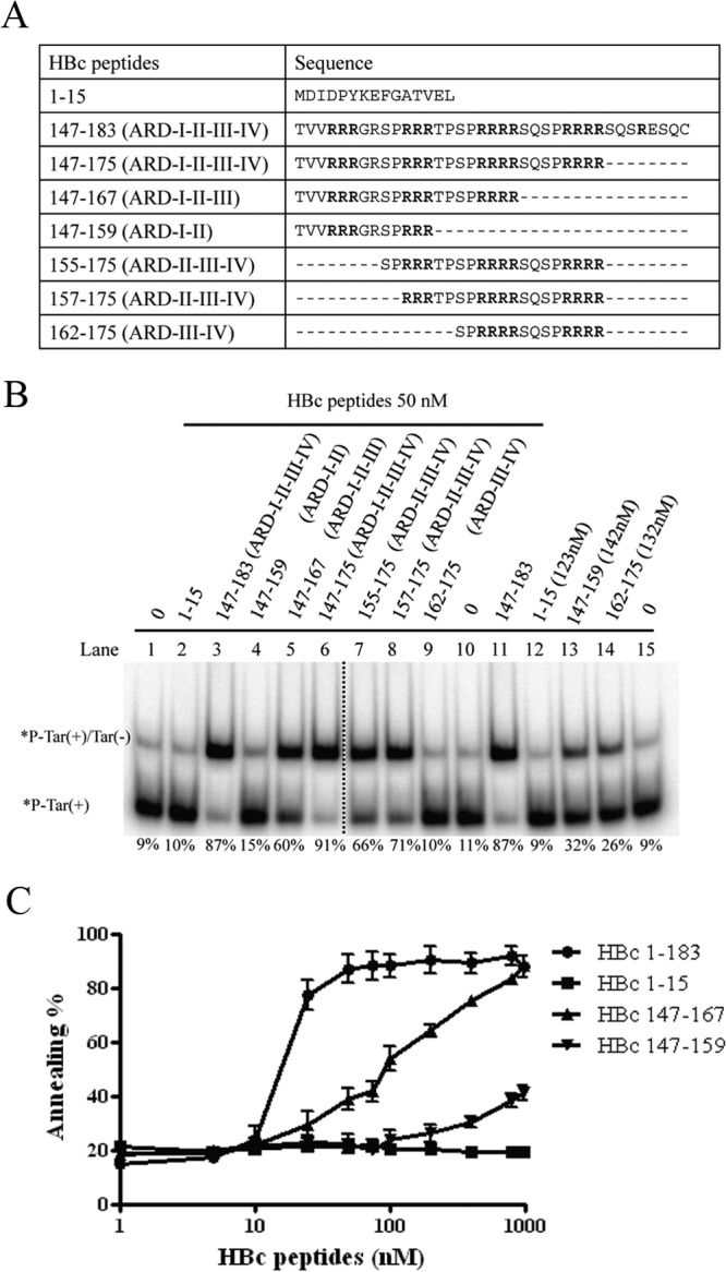
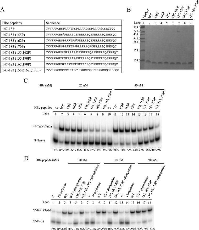
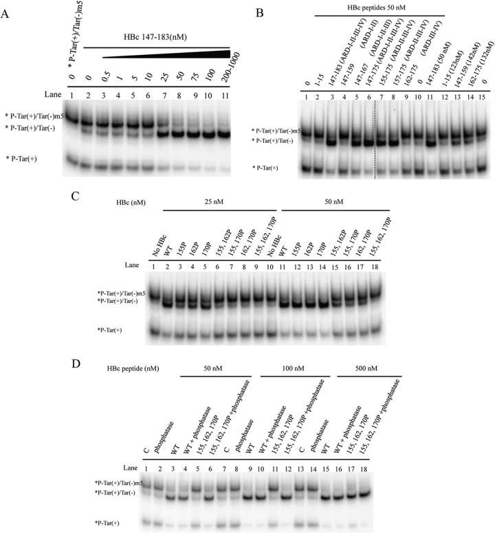
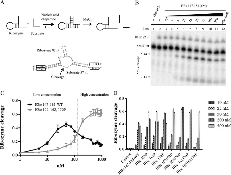
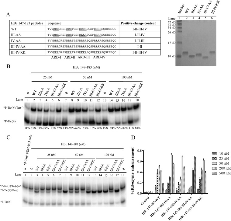
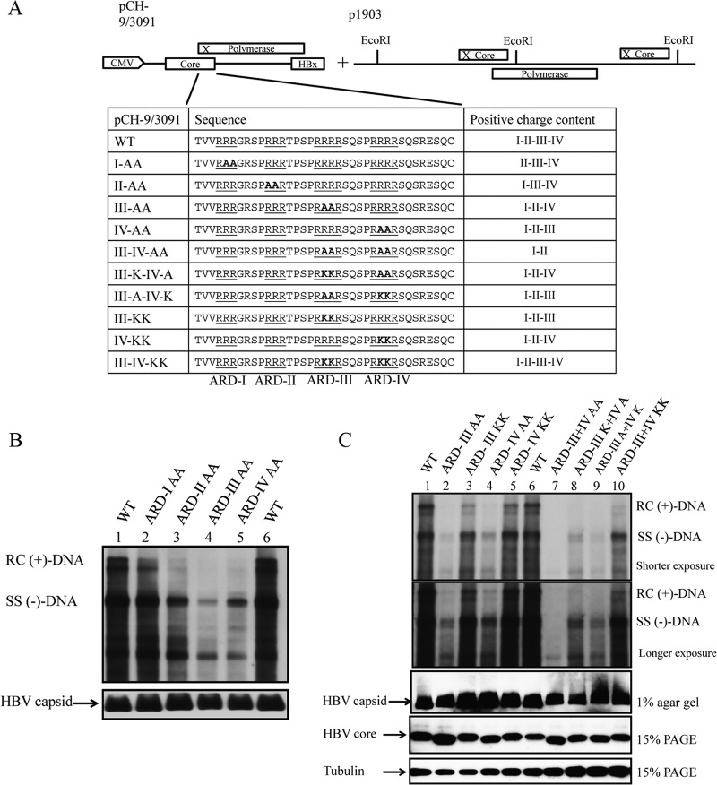
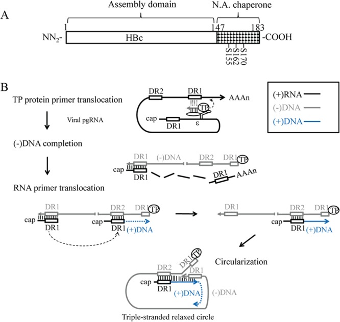
Hepatitis B virus (HBV) DNA replication occurs within the HBV icosahedral core particles. HBV core protein (HBc) contains an arginine-rich domain (ARD) at its carboxyl terminus. This ARD domain of HBc 149-183 is known to be important for viral replication but not known to have a structure. Recently, nucleocapsid proteins of several viruses have been shown to contain nucleic acid chaperone activity, which can facilitate structural rearrangement of viral genome. Major features of nucleic acid chaperones include highly basic amino acid residues and flexible protein structure. To test the nucleic acid chaperone hypothesis for HBc ARD, we first used the disassembled full-length HBc from Escherichia coli to analyze the nucleic acid annealing and strand displacement activities. To exclude the potential contamination of chaperones from E. coli, we designed synthetic HBc ARD peptides with different lengths and serine phosphorylations. We demonstrated that HBc ARD peptide can behave like a bona fide nucleic acid chaperone and that the chaperone activity depends on basic residues of the ARD domain. The loss of chaperone activity by arginine-to-alanine substitutions in the ARD can be rescued by restoring basic residues in the ARD. Furthermore, the chaperone activity is subject to regulation by phosphorylation and dephosphorylation at the HBc ARD. Interestingly, the HBc ARD can enhance in vitro cleavage activity of RNA substrate by a hammerhead ribozyme. We discuss here the potential significance of the HBc ARD chaperone activity in the context of viral DNA replication, in particular, at the steps of primer translocations and circularization of linear replicative intermediates.

Importance: Hepatitis B virus is a major human pathogen. At present, no effective treatment can completely eradicate the virus from patients with chronic hepatitis B. We report here a novel chaperone activity associated with the viral core protein. Our discovery could lead to a new drug design for more effective treatment against hepatitis B virus in the future.


Figures

FIG 1
FIG 1
Full-length HBc 1-183 of disassembled HBc particles can promote DNA annealing and strand displacement activities. (A) Schematic presentation of induction of capsid disassembly by treating HBc particles with nuclease S7 and low salt (29). According to the charge balance hypothesis (29, 32, 62), capsid instability and disassembly can be induced by the loss of encapsidated nucleic acid macromolecules upon nuclease S7 digestion. Capsid particle assembly and disassembly can be visualized in native agarose gel electrophoresis by SYPRO Ruby staining for protein or by SYBR green II staining for encapsidated RNA (29). Disassembled capsid protein monomers can be monitored in denaturing SDS-PAGE by Western blotting. (B) Encapsidated RNA of HBV capsids on a native agarose gel was stained with SYBR green II, and the same gel was later stained with SYPRO Ruby for HBc protein. No RNA or HBc protein signals were detected after treatment with micrococcal nuclease S7. Lower panel, full-length HBc protein monomer was detected by SDS-PAGE and Western blot analysis, indicating that HBc protein remains intact and full length after S7 treatment. (C) Cartoon illustration of the nucleic acid annealing assay. Radioactively labeled Tar(+) DNA and the complementary Tar(−) DNA oligonucleotides were incubated in the presence of HBc particles with or without capsid disassembly pretreatment. Efficient formation of the Tar(+)/Tar(−) duplex was detected only in the presence of disassembled particles. The oligonucleotides used in the experiment are indicated. (D) The kinetics of DNA annealing activity in the presence of assembled versus disassembled HBc particles were measured. A time course from 15 s to 5 min was conducted by using the Tar(−) and Tar(+) DNA annealing assay as detailed for panel C. (E) Graphic comparison of DNA annealing activity between assembled and disassembled HBc particles based on the data in panel D. The annealing activity (%) was calculated as the ratio of the banding intensities between duplex molecules and total intensities: [32P-labeled Tar(+)/Tar(−)]/{[32P-labeled Tar(+)/Tar(−)] + 32P-labeled Tar(+)}. (F) Schematic presentation of the DNA strand exchange assay. Radioactively labeled Tar(+) DNA was preannealed to the complementary Tar(−)m5 DNA, which contained a 5-nucleotide mismatch. Perfectly matched complementary Tar(−) DNA was added in the absence or presence of HBc ARD peptides. The oligonucleotides used in the experiment are indicated. (G) The kinetics of strand exchange (displacement) activity in the presence of assembled versus disassembled HBc particles were measured. A time course from 15 s to 5 min was conducted by using the Tar(−) and Tar(+) strand exchange assay as detailed for panel F.
FIG 2
FIG 2
Prediction of disorder in HBV core protein (HBc). (A) Full-length HBc contains a capsid assembly domain and an arginine-rich domain (ARD). Here we asked whether the nucleic acid chaperone activity of HBc could reside in the ARD domain. (B) Disordered regions in HBV core protein (accession number J02203) were predicted using the Protein DisOrder prediction system (http://prdos.hgc.jp/cgi-bin/top.cgi). If the amino acid score is over 0.5, this amino acid is considered to reside in a disordered environment. If the amino acid score is less than 0.5, this amino acid is within an ordered region. (C) The disordered residues are displayed in boldface, and four arginine-rich subdomains (ARD I to IV) are included in this region.
FIG 3
FIG 3
The arginine-rich domain (ARD) of HBc 147-183 exhibits DNA annealing activity. (A) DNA annealing activity is dependent on HBc ARD peptides in a dose-dependent manner. Annealing of Tar(+)/Tar(−) was promoted by HBc ARD peptides. 32P-labeled Tar(+) DNA (1 nM) was incubated with unlabeled Tar(−) DNA (1 nM) in the presence of increasing amounts of HBV core peptides HBc 147-183 (0.5 to 1000 nM) at 37°C for 5 min. (B) The annealing reaction is plotted against HBc ARD peptide concentrations based on the data in panel A. The reaction kinetics appeared to be cooperative after 10 nM before reaching a plateau. The annealing activity (percent) was calculated as the ratio of the banding intensities between duplex molecules and total intensities: [32P-labeled Tar(+)/Tar(−)]/{[32P-labeled Tar(+)/Tar(−)] + 32P-labeled Tar(+)}.
FIG 4
FIG 4
Mapping the minimal essential region of HBc ARD peptides required for DNA annealing activity. (A) Amino acid sequences of HBc ARD peptides used in the deletion mapping experiment. (B) Labeled Tar(+) and cold Tar(−) were incubated with HBc ARD peptides with different lengths and arginine contents. The dotted line between lanes 6 and 7 indicates that lanes 1 to 6 and lanes 7 to 15 are spliced from the same gel. (C) Dose-response curves for DNA annealing activities were compared by using various HBc ARD peptides with different lengths and arginine contents. The calculation of the efficiency of DNA annealing activity was as described for Fig. 1. As shown in panel A, peptides HBc 1-15, 147-159, 147-167, and 147-183 contain 0, 2, 3, and 4 ARD subdomains, respectively.
FIG 5
FIG 5
Phosphorylation and dephosphorylation of HBc ARD peptides can influence DNA annealing activity. (A) Amino acid sequences of different HBc ARD peptides phosphorylated at different positions. (B) Characterization of HBc ARD peptides by Tricine SDS-PAGE. Peptides were stained with Green Angel. (C) Labeled Tar(+) and cold Tar(−) were incubated with 25 and 50 nM phosphorylated HBc ARD peptides at 37°C for 5 min. (D) Rescue of the reduced DNA annealing activity of HBc ARD peptides by dephosphorylation treatment with lambda phosphatase.
FIG 6
FIG 6
Strand exchange (displacement) activities of HBc ARD peptides. (A) A DNA duplex of 32P-Tar(+)/Tar(−)m5 (1 nM) was incubated with an excessive amount of Tar(−) DNA (2 nM) in the absence or presence of HBc ARD peptides at various concentrations at 37°C for 5 min. (B) Deletion mapping of the minimal essential region of HBc peptides important for strand displacement activity. (C) Serine phosphorylation can attenuate the strand displacement activity of HBc ARD peptides. (D) Dephosphorylation of HBc ARD peptides by lambda phosphatase treatment can restore the chaperone activity of HBc ARD peptides in the strand displacement assay.
FIG 7
FIG 7
Enhancement of hammerhead ribozyme (HHR) cleavage by HBc ARD peptides. (A) Schematic diagrams of HHR and substrate 15bs RNAs. Only parts of the sequences of HHR and substrate 15bs are shown, and the arrow represents the cleavage point. (B) HBc ARD peptides promote hammerhead ribozyme cleavage. HHR RNA ribozyme and 15bs substrate RNA were incubated with various concentrations of HBc ARD peptides and analyzed as described in Materials and Methods. (C) Serine phosphorylation of HBc ARD peptides at lower concentrations attenuate the hammerhead RNA ribozyme cleavage activity yet increase the ribozyme activity at higher concentrations (above 120 nM). The cleavage activity of hammerhead ribozyme was calculated as the banding intensities of 44-nt and 13-nt cleaved substrates divided by the total intensities of 57-nt uncleaved substrates plus 44-nt and 13-nt cleaved substrates. (D) Hammerhead ribozyme cleavage activity using various phosphorylated ARD HBc 147-183 peptides.
FIG 8
FIG 8
Positive charge content of HBc ARD is important for nucleic acid chaperone activity. (A) Amino acid sequences of mutant HBc ARD peptides containing various arginine-to-alanine (R-to-A) or arginine-to-lysine (R-to-K) substitutions and characterization of HBc ARD peptides by Tricine SDS-PAGE. These peptides were stained with Green Angel. (B) The DNA annealing activity of HBc ARD peptides was significantly reduced by R-to-A substitutions (lanes 3 to 5 and 9 to 11) but not by R-to-K substitutions (lanes 6, 12, and 18). (C) The strand displacement activity of HBc ARD peptides was significantly reduced by R-to-A substitutions (lanes 4 to 6 and 9 to 11) but not by R-to-K substitutions (lanes 7, 12, and 17). (D) The enhancement of hammerhead ribozyme cleavage activity by HBc ARD peptides was significantly reduced by R-to-A substitutions, but not by R-to-K substitutions, at 25 and 50 nM.
FIG 9
FIG 9
Positive charge content of HBc ARD is important for HBV replication in Southern blot analysis. (A) Complementation analysis of HBV DNA replication was conducted by cotransfection of HBV replicon plasmid pCH-9/3091 containing R-to-A or R-to-K substitutions at the HBc ARD and core-deficient HBV tandem dimer plasmid p1903, which can provide functional polymerase. The amino acid sequences of HBc ARD mutations are designed to contain various positive charge contents by R-to-A or R-to-K substitutions. I to IV indicate the four different ARD subdomains. (B) HBV DNA replication of ARD mutants ARD-I to -IV in Huh7 cells was analyzed by Southern blotting assay. The most severe defect in replication was observed in HBc ARD-IIIAA and ARD-IVAA. The lower panel shows the control of intracellular HBV capsids from transfected cell lysates separated on 1% agarose gel and visualized by Western blotting using anti-HBc antibody. (C) However, the severe replication defect in mutant ARD-III AA and mutant ARD-IV AA was not observed for R-to-K substitutions in ARD-III and ARD-IV. Similarly, mutant ARD-III+IV AA, missing four arginines, is replication defective, yet no severe replication defect was observed for R-to-K substitutions in both ARD-III and ARD-IV. The lower panels (HBV capsids, HBV core protein, and tubulin) show controls for sample loading. This result is consistent with our previous studies of R-to-K substitutions in HBc ARD (32).
FIG 10
FIG 10
Nucleic acid chaperone hypothesis in HBV replication (A) Assignment of the nucleic acid (N.A.) chaperone activity to the ARD of HBc 147-183. Serines 155, 162, and 170 are subject to phosphorylation and dephosphorylation, which can modulate chaperone activity and viral replication. (B) A diagram of HBV DNA replication illustrates the specific steps where an HBc chaperone might play a role in nucleic acid structural rearrangement, such as annealing, unwinding, and strand displacement. Specifically, we hypothesize here that the HBc chaperone activity could facilitate TP protein primer translocation, which is important for HBV minus-strand DNA synthesis. In addition, it could facilitate RNA primer translocation, which is important for the initiation of plus-strand DNA synthesis. Finally, during the circularization of the linear minus-strand DNA, the 3′ DR1 could undergo a strand invasion and displacement of the 5′ DR1 sequences. Further details are as follows. The encapsidation signal ε is for TP protein binding. The two direct repeat sequences DR1 are located at the viral pgRNA 5′ and 3′-end regions, and the relative position of DR2 is also indicated. The viral pgRNA is associated with polymerase P protein and packaged into capsids during assembly. (i) For TP protein primer translocation, TP protein primes on the bulge of ε to initiate minus-strand synthesis. The nascent minus-strand DNA then translocates to the 3′ copy of DR1. (ii) The minus-strand DNA is elongated, and the pgRNA is degraded simultaneously by RNase H domain of polymerase. The 5′-end sequences of pgRNA, including the DR1, are somehow spared from RNase H degradation. (iii) For RNA primer translocation, this RNase H-spared 5′ pgRNA sequence could undergo a translocation from DR1 to DR2 and prime plus-strand DNA synthesis from DR2. (iv) For circularization, after the plus-strand DNA synthesis reaches the 5′ end of the minus-strand template, the 3′-end DR1 of the minus strand DNA could undergo a strand displacement of the 5′-end DR1 and form a relaxed-circle (RC) structure with a triple-strand region (53).

Similar articles

Cited by

References

    1. Purcell RH. 1994. Hepatitis viruses: changing patterns of human disease. Proc. Natl. Acad. Sci. U. S. A. 91:2401–2406. 10.1073/pnas.91.7.2401 - DOI - PMC - PubMed
    1. Shih C. 2012. Chronic hepatitis B and C: basic science to clinical applications. World Scientific, Hackensack, NJ
    1. Wieland SF, Chisari FV. 2005. Stealth and cunning: hepatitis B and hepatitis C viruses. J. Virol. 79:9369–9380. 10.1128/JVI.79.15.9369-9380.2005 - DOI - PMC - PubMed
    1. Seeger C, Mason WS. 2000. Hepatitis B virus biology. Microbiol. Mol. Biol. Rev. 64:51–68. 10.1128/MMBR.64.1.51-68.2000 - DOI - PMC - PubMed
    1. Newman M, Suk FM, Cajimat M, Chua PK, Shih C. 2003. Stability and morphology comparisons of self-assembled virus-like particles from wild-type and mutant human hepatitis B virus capsid proteins. J. Virol. 77:12950–12960. 10.1128/JVI.77.24.12950-12960.2003 - DOI - PMC - PubMed

Publication types

MeSH terms

LinkOut - more resources