. 2014 Mar;88(5):2572-83.
doi: 10.1128/JVI.02573-13. Epub 2013 Dec 18.

An ITAM in a nonenveloped virus regulates activation of NF-κB, induction of beta interferon, and viral spread

Affiliations

An ITAM in a nonenveloped virus regulates activation of NF-κB, induction of beta interferon, and viral spread

Rachael E Stebbing et al. J Virol. 2014 Mar.

Abstract

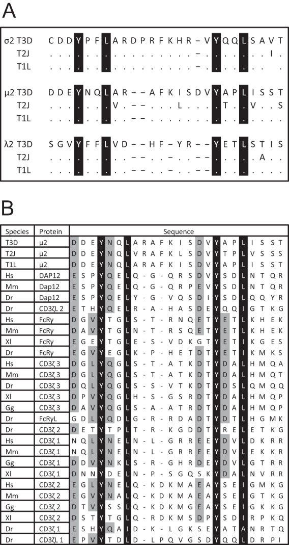
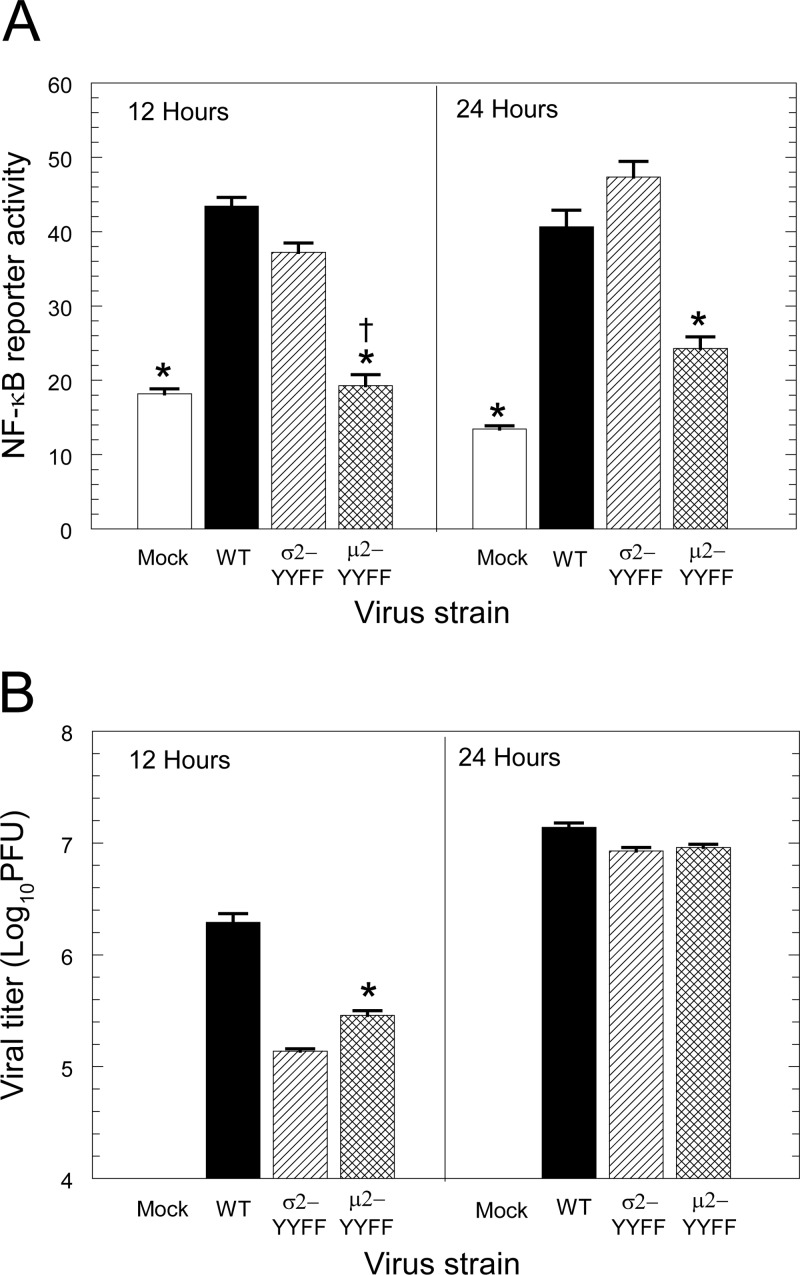
Immunoreceptor tyrosine-based activation motifs (ITAMs) are signaling domains located within the cytoplasmic tails of many transmembrane receptors and associated adaptor proteins that mediate immune cell activation. ITAMs also have been identified in the cytoplasmic tails of some enveloped virus glycoproteins. Here, we identified ITAM sequences in three mammalian reovirus proteins: μ2, σ2, and λ2. We demonstrate for the first time that μ2 is phosphorylated, contains a functional ITAM, and activates NF-κB. Specifically, μ2 and μNS recruit the ITAM-signaling intermediate Syk to cytoplasmic viral factories and this recruitment requires the μ2 ITAM. Moreover, both the μ2 ITAM and Syk are required for maximal μ2 activation of NF-κB. A mutant virus lacking the μ2 ITAM activates NF-κB less efficiently and induces lower levels of the downstream antiviral cytokine beta interferon (IFN-β) than does wild-type virus despite similar replication. Notably, the consequences of these μ2 ITAM effects are cell type specific. In fibroblasts where NF-κB is required for reovirus-induced apoptosis, the μ2 ITAM is advantageous for viral spread and enhances viral fitness. Conversely, in cardiac myocytes where the IFN response is critical for antiviral protection and NF-κB is not required for apoptosis, the μ2 ITAM stimulates cellular defense mechanisms and diminishes viral fitness. Together, these results suggest that the cell type-specific effect of the μ2 ITAM on viral spread reflects the cell type-specific effects of NF-κB and IFN-β. This first demonstration of a functional ITAM in a nonenveloped virus presents a new mechanism for viral ITAM-mediated signaling with likely organ-specific consequences in the host.


Figures

FIG 1
FIG 1
ITAM sequences are present in three reovirus proteins. (A) ITAMs in the reovirus σ2 (residues 127 to 151), μ2 (residues 115 to 138), and λ2 proteins (residues 668 to 688). ITAMs of prototype reovirus strains T3D, T2J, and T1L are shown. Sequences are from references , , and . Dots indicate identical residues and dashes indicate gaps in the alignment. (B) Reovirus μ2 ITAMs aligned with cellular ITAMs. Black indicates conserved ITAM residues. Gray indicates additional residues common to many ITAMs. Hs, Homo sapiens (human); Mm, Mus musculus (mouse); Dr, Danio rerio (zebrafish); Xl, Xenopus laevis; Gg, Gallus gallus (chicken). The extensions 1, 2, and 3 following protein names distinguish between the three ITAMs in each of the indicated proteins.
FIG 2
FIG 2
μ2 is phosphorylated on tyrosine residues. AD-293 cells were transfected with the indicated FLAG-tagged plasmid. At 40 h posttransfection, whole-cell lysates were immunoprecipitated using anti-FLAG beads, and fractions from duplicate samples were resolved by SDS-PAGE and immunoblotted with anti-FLAG or anti-phosphotyrosine antibodies. The results are representative of at least two independent experiments.
FIG 3
FIG 3
NF-κB is activated by μ2 and maximal activation requires the μ2 ITAM. (A) HEK-293 cells were cotransfected with the indicated FLAG-tagged plasmid, a constitutively expressing Renilla luciferase plasmid for normalizations, and a luciferase reporter plasmid regulated by NF-κB, IFN-β, or ISRE promoters. At 24 h posttransfection, luciferase activity was quantified. The results are expressed as means ± the standard errors of the mean of five replicate samples and are representative of two independent experiments. (B) Whole-cell lysates corresponding to panel A were resolved by SDS-PAGE, transferred to a nitrocellulose membrane, and immunoblotted with anti-FLAG or anti-GAPDH antibodies. Gels were scanned, and band intensities were quantified. The results are presented as the FLAG intensity normalized to the GAPDH intensity and are representative of at least two independent experiments. *, Significantly different from pFLAG and pFLAG-μ2-YYFF (P < 0.05).
FIG 4
FIG 4
μ2 requires Syk for maximal activation of NF-κB. (A) AD-293 cells were treated for 1 h with DMSO alone or 25 μM the Syk-specific inhibitor piceatannol and then cotransfected with the indicated effector plasmid, a constitutively expressing Renilla luciferase plasmid for normalizations, and an NF-κB-luciferase reporter plasmid. At 24 h posttransfection, the luciferase activity was quantified. The results are expressed as means plus the standard errors of the mean of six replicate samples and are representative of at least two independent experiments. (B) The indicated amount (in μg) of whole-cell lysates corresponding to panel A were resolved by SDS-PAGE, transferred to a nitrocellulose membrane, and immunoblotted with anti-μ2 or anti-actin-HRP antibodies. The results are representative of at least two independent experiments. *, Significantly different from pCAGGS-μ2 and pFLAG-μ2 in piceatannol and pFLAG in either treatment (P < 0.02).
FIG 5
FIG 5
μ2 and μNS recruit Syk to viral factories and recruitment requires the μ2 ITAM. Vero cells were cotransfected with the indicated plasmids. At 24 h posttransfection, the cells were fixed and immunostained, and nuclei were stained with DAPI. (A) Cells were transfected with GFP-μNS (green) and the indicated μ2-expressing plasmid and then immunostained with anti-μ2 antibody (red). (B) Cells were transfected as in panel A and immunostained with anti-μ2 (green) and anti-tubulin (red) antibodies. (C) Cells were transfected as in panel A with the addition of a Syk-expressing plasmid and immunostained with anti-Syk (red) and anti-μ2 (green) antibodies. The results are representative of three independent experiments. Scale bar, 20 μm; inset scale bar, 10 μm.
FIG 6
FIG 6
The μ2 ITAM regulates activation of NF-κB during viral infection. (A) AD-293 cells were transfected with an NF-κB-luciferase reporter plasmid and a constitutively expressing Renilla luciferase plasmid for normalizations. At 24 h posttransfection, cells were infected with the indicated virus at an MOI of 10 PFU per cell. At 12 and 24 h postinfection, the luciferase activity was quantified. The results are expressed as means ± the standard deviations of four or five replicate wells and are representative of at least three independent experiments. (B) AD-293 cells were infected with the indicated virus at an MOI of 3 PFU per cell. At 12 and 24 h postinfection, the virus titers were determined by plaque assay. The results are expressed as the means of quadruplicate samples plus the standard deviations for a representative of at least two independent experiments. *, Significantly different from WT T3D and σ2-YYFF (P < 0.05); †, not significantly different from mock (P > 0.05).
FIG 7
FIG 7
The μ2 ITAM regulates induction of IFN-β during viral infection. (A) L929 cells or primary cardiac myocyte cultures were infected at an MOI of 25 or 10 PFU per cell, respectively. At 8 or 12 h postinfection, RNA was quantified by reverse transcription and quantitative real-time PCR, and the copy number was normalized to GAPDH. The fold induction of IFN-β is expressed relative to mock-infected cultures. The results are expressed as means ± the standard errors of the mean of triplicate wells for a representative of three independent experiments. (B) L929 cells or primary cardiac myocytes cultures were infected with the indicated virus at an MOI of 3 PFU per cell. At 8 h postinfection, the virus titers were determined by plaque assay. The results are expressed as means plus the standard deviations for two or three independent experiments each with triplicate wells. *, Significantly different from WT T3D and σ2-YYFF (P < 0.05); **, significantly different from σ2-YYFF (P < 0.05).
FIG 8
FIG 8
The effect of the μ2 ITAM on virus titer after multiple cycles of replication is cell type specific and was determined by the IFN-β response. (A) L929 cells and primary cardiac myocyte cultures were infected with the indicated virus at an MOI of 0.1 PFU per cell. At 5 days postinfection, the virus titers were determined by plaque assay. The results are expressed as means ± the standard deviations of triplicate wells for a representative of at least two independent experiments. (B) L929 cells and primary cardiac myocyte cultures were infected at an MOI of 0.1 or 0.3 PFU per cell, respectively. At 1 h postinfection, inocula were removed and replaced with either medium alone or medium containing anti-IFN-β antibody. At 2 days postinfection, additional anti-IFN-β antibody was added to appropriate wells. The results are expressed as the means of quadruplicate samples plus the standard deviations for a representative of at least two independent experiments. *, Significantly different from WT T3D and σ2-YYFF (P < 0.05).
FIG 9
FIG 9
The effect of the μ2 ITAM on cytopathic effect after multiple cycles of replication is cell type specific. L929 cells or primary cardiac myocyte cultures were infected with the indicated virus at an MOI of 0.1 or 1 PFU/cell, respectively (the cytopathic effect was undetectable in cardiac myocytes infected at 0.1 PFU/cell; data not shown). At 5 and 7 days postinfection, the cell viability was quantified by using an MTT assay. The results from a minimum of four replicate wells per infection are expressed as the means plus the standard errors of the mean relative to mock-infected cultures for a representative of at least two independent experiments. *, Significantly different from WT T3D and σ2-YYFF (P < 0.05).

Similar articles

Cited by

References

    1. Underhill DM, Goodridge HS. 2007. The many faces of ITAMs. Trends Immunol. 28:66–73. 10.1016/j.it.2006.12.004 - DOI - PubMed
    1. Berton G, Mocsai A, Lowell CA. 2005. Src and Syk kinases: key regulators of phagocytic cell activation. Trends Immunol. 26:208–214. 10.1016/j.it.2005.02.002 - DOI - PubMed
    1. Mocsai A, Ruland J, Tybulewicz VL. 2010. The SYK tyrosine kinase: a crucial player in diverse biological functions. Nat. Rev. Immunol. 10:387–402. 10.1038/nri2765 - DOI - PMC - PubMed
    1. Sada K, Takano T, Yanagi S, Yamamura H. 2001. Structure and function of Syk protein-tyrosine kinase. J. Biochem. 130:177–186. 10.1093/oxfordjournals.jbchem.a002970 - DOI - PubMed
    1. Barrow AD, Trowsdale J. 2006. You say ITAM and I say ITIM, let's call the whole thing off: the ambiguity of immunoreceptor signaling. Eur. J. Immunol. 36:1646–1653. 10.1002/eji.200636195 - DOI - PubMed

Publication types

MeSH terms

LinkOut - more resources