Interaction of foot-and-mouth disease virus nonstructural protein 3A with host protein DCTN3 is important for viral virulence in cattle
- PMID: 24352458
- PMCID: PMC3958104
- DOI: 10.1128/JVI.03059-13
Interaction of foot-and-mouth disease virus nonstructural protein 3A with host protein DCTN3 is important for viral virulence in cattle
Abstract
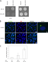
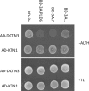
Nonstructural protein 3A of foot-and-mouth disease virus (FMDV) is a partially conserved protein of 153 amino acids in most FMDVs examined to date. The role of 3A in virus growth and virulence within the natural host is not well understood. Using a yeast two-hybrid approach, we identified cellular protein DCTN3 as a specific host binding partner for 3A. DCTN3 is a subunit of the dynactin complex, a cofactor for dynein, a motor protein. The dynactin-dynein duplex has been implicated in several subcellular functions involving intracellular organelle transport. The 3A-DCTN3 interaction identified by the yeast two-hybrid approach was further confirmed in mammalian cells. Overexpression of DCTN3 or proteins known to disrupt dynein, p150/Glued and 50/dynamitin, resulted in decreased FMDV replication in infected cells. We mapped the critical amino acid residues in the 3A protein that mediate the protein interaction with DCTN3 by mutational analysis and, based on that information, we developed a mutant harboring the same mutations in O1 Campos FMDV (O1C3A-PLDGv). Although O1C3A-PLDGv FMDV and its parental virus (O1Cv) grew equally well in LFBK-αvβ6, O1C3A-PLDGv virus exhibited a decreased ability to replicate in primary bovine cell cultures. Importantly, O1C3A-PLDGv virus exhibited a delayed disease in cattle compared to the virulent parental O1Campus (O1Cv). Virus isolated from lesions of animals inoculated with O1C3A-PLDGv virus contained amino acid substitutions in the area of 3A mediating binding to DCTN3. Importantly, 3A protein harboring similar amino acid substitutions regained interaction with DCTN3, supporting the hypothesis that DCTN3 interaction likely contributes to virulence in cattle.
Importance: The objective of this study was to understand the possible role of a FMD virus protein 3A, in causing disease in cattle. We have found that the cellular protein, DCTN3, is a specific binding partner for 3A. It was shown that manipulation of DCTN3 has a profound effect in virus replication. We developed a FMDV mutant virus that could not bind DCTN3. This mutant virus exhibited a delayed disease in cattle compared to the parental strain highlighting the role of the 3A-DCTN3 interaction in virulence in cattle. Interestingly, virus isolated from lesions of animals inoculated with mutant virus contained mutations in the area of 3A that allowed binding to DCTN3. This highlights the importance of the 3A-DCTN3 interaction in FMD virus virulence and provides possible mechanisms of virus attenuation for the development of improved FMD vaccines.
Figures


Similar articles
-
Cellular Vimentin Interacts with Foot-and-Mouth Disease Virus Nonstructural Protein 3A and Negatively Modulates Viral Replication.J Virol. 2020 Jul 30;94(16):e00273-20. doi: 10.1128/JVI.00273-20. Print 2020 Jul 30. J Virol. 2020. PMID: 32493819 Free PMC article.
-
Substitution 3A protein of foot-and-mouth disease virus of attenuated ZB strain rescued the viral replication and infection in bovine cells.Res Vet Sci. 2020 Feb;128:145-152. doi: 10.1016/j.rvsc.2019.11.001. Epub 2019 Nov 6. Res Vet Sci. 2020. PMID: 31791012
-
Genome sequence of foot-and-mouth disease virus outside the 3A region is also responsible for virus replication in bovine cells.Virus Res. 2016 Jul 15;220:64-9. doi: 10.1016/j.virusres.2016.04.011. Epub 2016 Apr 16. Virus Res. 2016. PMID: 27094491
-
Construction and characterization of 3A-epitope-tagged foot-and-mouth disease virus.Infect Genet Evol. 2015 Apr;31:17-24. doi: 10.1016/j.meegid.2015.01.003. Epub 2015 Jan 10. Infect Genet Evol. 2015. PMID: 25584768
-
Global Foot-and-Mouth Disease Research Update and Gap Analysis: 7 - Pathogenesis and Molecular Biology.Transbound Emerg Dis. 2016 Jun;63 Suppl 1:63-71. doi: 10.1111/tbed.12520. Transbound Emerg Dis. 2016. PMID: 27320168 Review.
Cited by
-
Senecavirus A 3C Protease Mediates Host Cell Apoptosis Late in Infection.Front Immunol. 2019 Mar 13;10:363. doi: 10.3389/fimmu.2019.00363. eCollection 2019. Front Immunol. 2019. PMID: 30918505 Free PMC article.
-
Foot-and-mouth disease virus type O specific mutations determine RNA-dependent RNA polymerase fidelity and virus attenuation.Virology. 2018 May;518:87-94. doi: 10.1016/j.virol.2018.01.030. Epub 2018 Feb 20. Virology. 2018. PMID: 29455065 Free PMC article.
-
Porcine Picornavirus 3C Protease Degrades PRDX6 to Impair PRDX6-mediated Antiviral Function.Virol Sin. 2021 Oct;36(5):948-957. doi: 10.1007/s12250-021-00352-4. Epub 2021 Mar 15. Virol Sin. 2021. PMID: 33721217 Free PMC article.
-
Foot-and-Mouth Disease Virus 3A Hijacks Sar1 and Sec12 for ER Remodeling in a COPII-Independent Manner.Viruses. 2022 Apr 18;14(4):839. doi: 10.3390/v14040839. Viruses. 2022. PMID: 35458569 Free PMC article.
-
Genome Sequencing of Foot-and-Mouth Disease Virus Type O Isolate GRE/23/94.Genome Announc. 2016 May 12;4(3):e00353-16. doi: 10.1128/genomeA.00353-16. Genome Announc. 2016. PMID: 27174269 Free PMC article.
References
-
- Rowlands DJ. 2003. Special issue—Foot-and-mouth disease: preface. Virus Res. 91:1. 10.1016/S0168-1702(02)00264-2 - DOI
MeSH terms
Substances
LinkOut - more resources
Full Text Sources
Other Literature Sources
Research Materials

