. 2014 Mar;88(5):2821-34.
doi: 10.1128/JVI.03126-13. Epub 2013 Dec 18.

Kaposi's sarcoma-associated herpesvirus induces the ATM and H2AX DNA damage response early during de novo infection of primary endothelial cells, which play roles in latency establishment

Affiliations

Kaposi's sarcoma-associated herpesvirus induces the ATM and H2AX DNA damage response early during de novo infection of primary endothelial cells, which play roles in latency establishment

Vivek Vikram Singh et al. J Virol. 2014 Mar.

Abstract

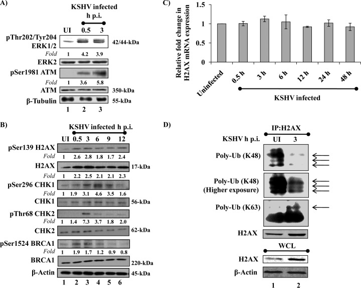
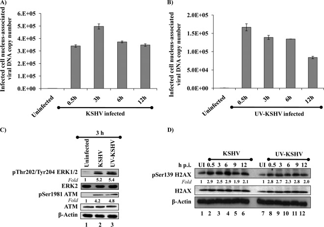
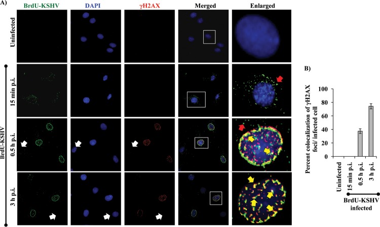
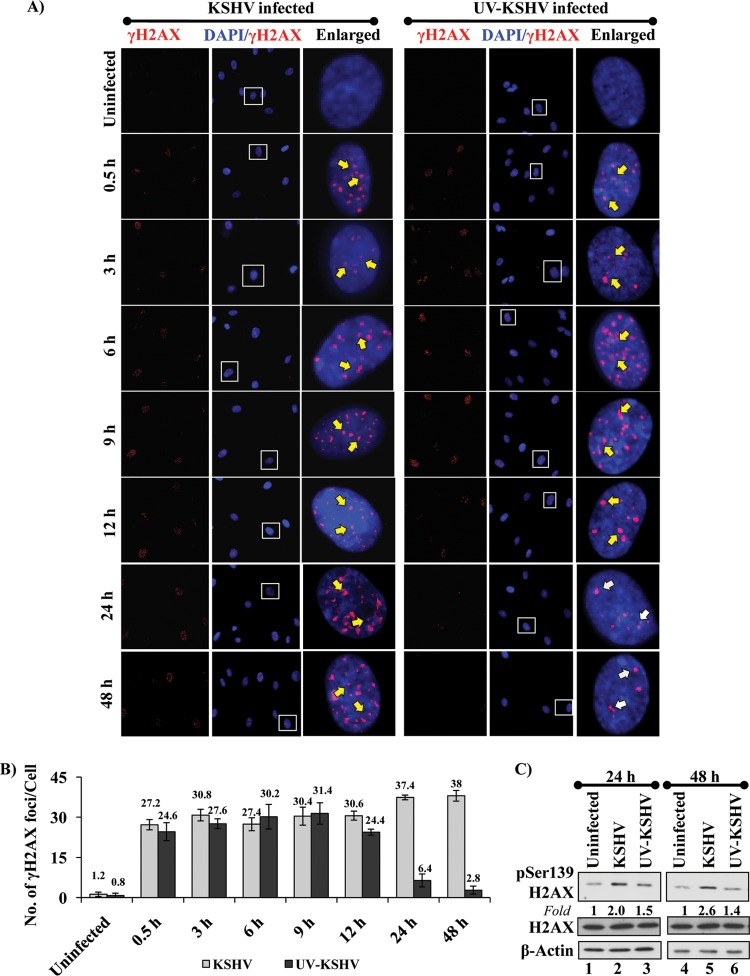
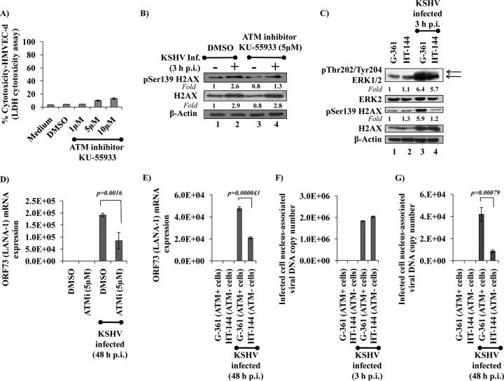
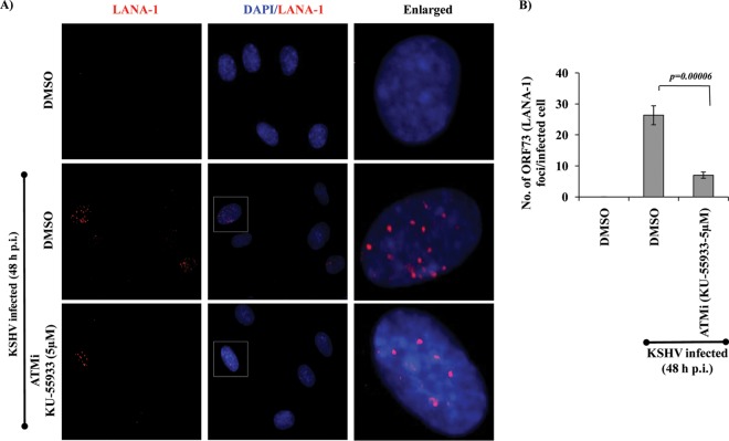
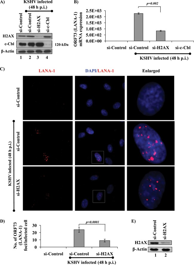
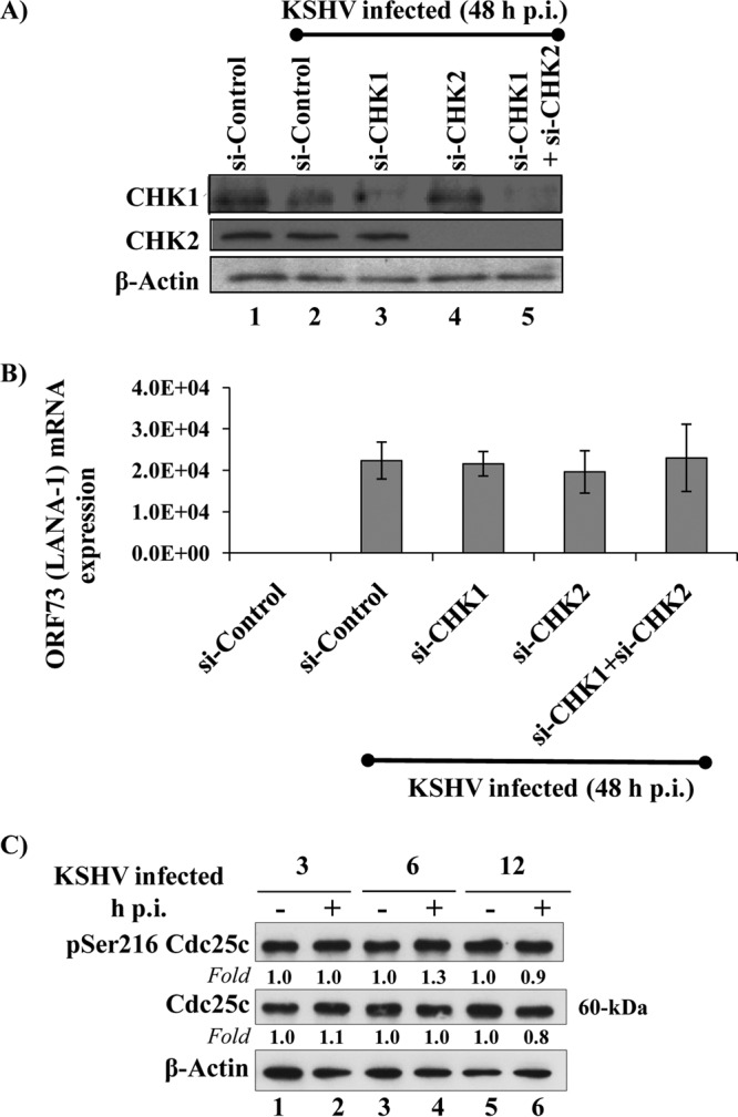
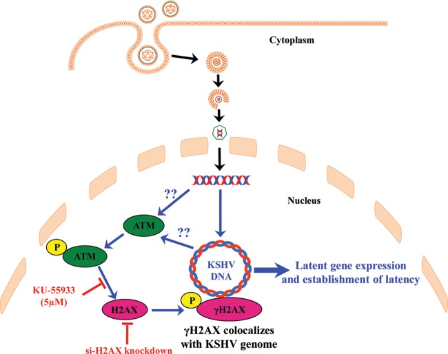
The DNA damage response (DDR) that evolved to repair host cell DNA damage also recognizes viral DNA entering the nucleus during infections. Here, we investigated the modulation of DDR signaling during de novo infection of primary endothelial cells by Kaposi's sarcoma-associated herpesvirus (KSHV). Phosphorylation of representative DDR-associated proteins, such as ataxia telangiectasia mutated (ATM) and H2AX, was induced as early as 30 min (0.5 h) postinfection and persisted during in vitro KSHV latency. Phosphorylated H2AX (γH2AX) colocalized at 30 min (0.5 h) with the KSHV genome entering the nuclei. Total H2AX protein levels also increased, and the increase was attributed to a decrease in degradative H2AX Lys48-linked polyubiquitination with a concomitant increase in Lys63-linked polyubiquitination that was shown to increase protein stability. ATM and H2AX phosphorylation and γH2AX nuclear foci were also induced by UV-inactivated KSHV, which ceased at later times of infection. Inhibition of ATM kinase activity by KU-55933 and H2AX knockdown by small interfering RNA significantly reduced the expression of the KSHV latency-associated nuclear antigen 1 (LANA-1; ORF73) and LANA-1 nuclear puncta. Knockdown of H2AX also resulted in a >80% reduction in the nuclear KSHV DNA copy numbers. Similar results were also observed in ATM-negative cells, although comparable levels of viral DNA entered ATM-negative and ATM-positive cell nuclei. In contrast, knockdown of CHK1 and CHK2 did not affect ORF73 expression. Collectively, these results demonstrate that KSHV induces ATM and H2AX, a selective arm of the DDR, for the establishment and maintenance of its latency during de novo infection of primary endothelial cells.

Importance: Eukaryotic cells mount a DNA damage response (DDR) to sense and repair different types of cellular DNA damage. In addition, DDR also recognizes exogenous genetic material, such as the viral DNA genome entering the nucleus during infections. The present study was undertaken to determine whether de novo Kaposi's sarcoma-associated herpesvirus (KSHV) infection modulates DDR. Our results demonstrate that early during de novo infection of primary endothelial cells, KSHV induces a selective arm of DDR signaling, such as the ATM kinase and its downstream target, H2AX, which are essential for KSHV's latent gene expression and the establishment of latency. These studies suggest that targeting ATM and H2AX could serve as an attractive strategy to block the establishment of KSHV latent infection and the associated malignancies.


Figures

FIG 1
FIG 1
Induction of DDR signaling and effect on the expression of H2AX mRNA during de novo KSHV infection of HMVEC-d. (A) Primary endothelial cells (HMVEC-d) infected with KSHV (30 DNA copies/cell) were analyzed for ERK1/2 and ATM phosphorylation and their total protein levels by immunoblot analysis. Fold changes were calculated by considering the levels for uninfected (UI) cells to be 1. (B) HMVEC-d infected with KSHV (30 DNA copies/cell) for various times at early stages of infection were analyzed for phosphorylation of H2AX, CHK1, CHK2, and BRCA1 along with their total protein levels by immunoblot analysis. β-Tubulin and β-actin were used as loading controls. Fold changes were calculated by considering the levels for uninfected cells to be 1. (C) Primary HMVEC-d infected with KSHV (30 DNA copies/cell) for various times were analyzed for H2AX gene expression by real-time RT-PCR. Each bar represents the fold increase in gene expression ± SD for three independent experiments. Fold changes were calculated by considering the levels for uninfected cells to be 1 after normalizing by the expression of the β-tubulin gene. (D) Primary HMVEC-d infected with KSHV for 3 h were analyzed for KSHV-induced polyubiquitination (Poly-Ub) of H2AX. Immunoprecipitation (IP) of 200 μg of the whole-cell lysate (WCL) was carried out with rabbit anti-H2AX antibodies, and samples were subjected to Western blot analysis for either Lys48- or Lys63-specific polyubiquitination. Blots were stripped and reprobed with H2AX. Arrows, polyubiquitination (first and second panels, K48; third panel, K63) of H2AX.
FIG 2
FIG 2
Induction of ATM and H2AX phosphorylation during de novo infection of HMVEC-d with live KSHV and UV-KSHV. (A and B) HMVEC-d infected with KSHV or UV-KSHV (30 DNA copies/cell) were analyzed for the kinetics of nuclear delivery of the KSHV genome by real-time DNA PCR of isolated nuclei from uninfected or infected cells. ORF73 standards and nontemplate controls were run in parallel. The viral DNA copy numbers were calculated from a standard graph generated by real-time DNA PCRs of known concentrations of a cloned ORF73 gene. Each reaction was done in triplicate, and each bar represents the mean ± SD for three experiments. (C) HMVEC-d infected with KSHV or UV-KSHV for 3 h were analyzed for phosphorylation of ERK1/2 and ATM along with their total protein levels by immunoblot analysis. Fold changes were calculated by considering the levels for uninfected cells to be 1. (D) HMVEC-d infected with KSHV or UV-KSHV for the indicated times were analyzed for phosphorylation of H2AX and total protein levels by immunoblot analysis. β-Actin was used as a loading control. Fold changes were calculated by considering the levels for uninfected cells to be 1.
FIG 3
FIG 3
Immunofluorescence analysis of γH2AX colocalization with the BrdU-labeled KSHV genome early during de novo infection of HMVEC-d. (A) HMVEC-d were infected with BrdU-labeled KSHV (30 DNA copies/cell) for 2 h, uninternalized virus was removed by washing, and infected and uninfected cells were further incubated at 37°C for the indicated times. The cells were washed, fixed, permeabilized, and blocked with Image-iT FX signal enhancer. Cells were stained with anti-BrdU and anti-γH2AX antibodies and visualized by incubation with Alexa 488 (green) and Alexa 594 (red) secondary antibodies, respectively. The image was merged with an image of DAPI (4′,6-diamidino-2-phenylindole)-stained nuclei. The boxed areas were enlarged and are presented in the rightmost panels. Yellow arrows, γH2AX foci colocalizing with the KSHV genome in infected cells; red arrows, the KSHV genome not internalized in the nucleus; white arrows, uninfected cells. Image results are depicted from a representative field taken from three independent experiments. Magnifications, ×60. (B) Percent colocalization of γH2AX foci per infected cell. A minimum of 3 fields having at least 15 cells was chosen, and the error bars show means ± SDs.
FIG 4
FIG 4
Immunofluorescence analysis of γH2AX levels early during de novo infection of HMVEC-d by live KSHV and UV-KSHV. (A) HMVEC-d were infected with KSHV (30 DNA copies/cell) for 2 h, uninternalized virus was removed by washing, and infected and uninfected cells were further incubated at 37°C for the indicated times. The cells were washed, fixed, permeabilized, and blocked with Image-iT FX signal enhancer. Cells were stained with anti-γH2AX antibodies and visualized by incubation with Alexa 594 (red) secondary antibodies. The image was merged with an image of DAPI-stained nuclei. The boxed areas were enlarged and are presented in the rightmost panels. Yellow arrows, γH2AX foci in KSHV-infected cells; white arrows, the decrease in γH2AX focus formation in UV-KSHV-infected cells. Image results are depicted from a representative field from three independent experiments. Magnifications, ×60. (B) The number of γH2AX foci per cell. A minimum of 3 fields having at least 15 cells was chosen, and the error bars show means ± SDs. (C) HMVEC-d infected with live KSHV or UV-KSHV (30 DNA copies/cell) for the indicated times were analyzed for phosphorylation of H2AX and total protein levels by immunoblot analysis. β-Actin was used as a loading control. Fold changes were calculated by considering the levels for uninfected cells to be 1.
FIG 5
FIG 5
Effect of ATM kinase activity inhibition by KU-55933 on KSHV-induced phosphorylation of H2AX and latent ORF73 gene expression during de novo infection of HMVEC-d. (A) Supernatants of HMVEC-d treated with DMSO or 1 μM, 5 μM, and 10 μM concentrations of an ATM kinase inhibitor for 2 h were analyzed for their effect on cellular viability by an LDH-based toxicity assay. (B) HMVEC-d pretreated with DMSO or an ATM kinase inhibitor for 2 h were infected (Inf.) with KSHV (30 DNA copies/cell). At 3 h postinfection, cellular lysates were analyzed for phosphorylation of H2AX and total protein levels by immunoblot analysis. Fold changes were calculated by considering the levels for uninfected cells to be 1. (C) ATM-positive G-361 (ATCC CRL-1424) and ATM-negative HT-144 (ATCC HTB-63) melanoma cells infected with KSHV (30 DNA copies/cell) were analyzed for ERK1/2 and H2AX phosphorylation and their total protein levels by immunoblot analysis. Fold changes were calculated by considering the levels for uninfected cells to be 1. (D) HMVEC-d treated with DMSO or an ATM kinase inhibitor (ATMi) for 2 h were infected with KSHV. At 48 h p.i., cells were harvested for RNA isolation and viral gene expression was determined by real-time RT-PCR with KSHV ORF73 gene-specific primers. Data represented are means ± SDs for three experiments. (E) ATM+ and ATM cells were infected with KSHV for 2 h. At 48 h p.i., cells were harvested for RNA isolation and viral gene expression was determined by real-time RT-PCR with KSHV ORF73 gene-specific primers. The data represented are means ± SDs for three experiments. (F and G) ATM-positive and -negative cells were infected with KSHV (30 DNA copies/cell) for 3 h (F) and 48 h (G). Nuclear delivery of the viral genome was determined by real-time DNA PCR using the isolated nuclei of uninfected or infected cells. Each reaction was done in triplicate, and each bar represents the mean ± SD for three experiments. The viral DNA copy numbers were calculated from a standard curve generated by real-time PCRs of known concentrations of a cloned ORF73 gene. Each reaction was done in triplicate, and each bar represents the mean ± SD for three experiments.
FIG 6
FIG 6
Effect of inhibition of ATM kinase activity by KU-55933 in latency establishment during de novo KSHV infection of HMVEC-d. (A) HMVEC-d treated with DMSO or an ATM kinase inhibitor for 2 h were infected with KSHV (30 DNA copies/cell), uninternalized virus was removed by washing, and infected and uninfected cells were further incubated at 37°C for 48 h. The cells were washed, fixed, permeabilized, and blocked with Image-iT FX signal enhancer. Cells were stained with anti-LANA-1 antibodies and visualized by incubation with Alexa 594 (red) secondary antibodies. The image was merged with images of DAPI-stained nuclei. The boxed areas were enlarged and are presented in the rightmost panels. Image results are depicted from a representative field taken from three independent experiments. Magnifications, ×60. (B) Number of characteristic LANA-1 puncta per infected cell. A minimum of 3 fields having at least 15 cells was chosen, and the error bars show means ± SDs.
FIG 7
FIG 7
Effect of H2AX knockdown on KSHV ORF73 gene expression and latency establishment during de novo infection of HMVEC-d. (A) The knockdown efficiency of si-control, si-H2AX, and si-c-Cbl transfection in HMVEC-d was determined by Western blotting. β-Actin was used as a loading control. (B) si-control-, si-H2AX-, or si-c-Cbl-transfected HMVEC-d were infected with KSHV for 2 h. At 48 h p.i., cells were harvested for RNA isolation and viral gene expression was determined by real-time RT-PCR with KSHV ORF73 gene-specific primers. Data represented are means ± SDs for three experiments. (C) si-control- and si-H2AX-transfected HMVEC-d were infected with KSHV (30 DNA copies/cell) for 2 h, uninternalized virus was removed by washing, and infected and uninfected cells were further incubated at 37°C for the indicated times. The cells were washed, fixed, permeabilized, and blocked with Image-iT FX signal enhancer. Cells were stained with anti-LANA-1 antibodies and visualized by incubation with Alexa 594 (red) secondary antibodies. The image was merged with images of DAPI-stained nuclei. The boxed areas were enlarged and are presented in the rightmost panels. Image results are depicted from a representative field taken from three independent experiments. Magnifications, ×60. (D) Number of characteristic LANA-1 puncta per infected cell. A minimum of 3 fields having at least 15 cells was chosen, and the error bars show means ± SDs. (E) Knockdown efficiency of si-H2AX transfection in HMVEC-d was determined by Western blotting. β-Actin was used as a loading control.
FIG 8
FIG 8
Effect of H2AX knockdown on kinetics of KSHV ORF73 expression and infected cell nucleus-associated viral DNA copy number during de novo infection of HMVEC-d. (A) The knockdown efficiency of si-H2AX in HMVEC-d was determined by Western blotting. β-Actin was used as a loading control. (B) The supernatants of nontransfected or mock-, si-control-, or si-H2AX-transfected HMVEC-d were analyzed 48 h after transfection for their effect on cellular viability by an LDH-based toxicity assay. (C) si-control- and si-H2AX-transfected HMVEC-d were infected with KSHV for 2 h. At the indicated times p.i., cells were harvested for RNA isolation and viral gene expression was determined by real-time RT-PCR with KSHV ORF73 gene-specific primers. Data represented are means ± SDs (n = 3). (D) si-control- and si-H2AX-transfected HMVEC-d were infected with KSHV (30 DNA copies/cell) for 48 h and analyzed for the KSHV genome associated with infected cell nuclei by real-time DNA PCR with ORF73 gene-specific primers. The viral DNA copy numbers were calculated from a standard graph generated by real-time PCRs of known concentrations of a cloned ORF73 gene. Each reaction was done in triplicate, and each bar represents the mean ± SD for three experiments.
FIG 9
FIG 9
Effect of CHK1 and CHK2 knockdown on KSHV ORF73 gene expression during de novo infection of HMVEC-d. (A) The knockdown efficiency of si-CHK1, si-CHK2, or si-CHK1 plus si-CHK2 transfection in HMVEC-d was determined by Western blotting. β-Actin was used as a loading control. (B) HMVEC-d transfected with si-control, si-CHK1, si-CHK2, or si-CHK1 plus si-CHK2 were infected with KSHV for 2 h. At 48 h p.i., cells were harvested for RNA isolation and viral gene expression was determined by real-time RT-PCR with KSHV ORF73 gene-specific primers. Data represented are means ± SDs for three experiments. (C) KSHV (30 DNA copies/cell)-infected HMVEC-d were analyzed for phosphorylated Cdc25c and total protein levels at different time points by immunoblot analysis. β-Actin was used as a loading control.
FIG 10
FIG 10
Schematic representation of the KSHV-induced DNA damage response (DDR) during de novo infection of endothelial cells and implications for the establishment of infection. During de novo KSHV infection of primary HMVEC-d, the linear dsDNA in the virions delivered into the nuclei as early as 30 min (0.5 h) postinfection circularizes. DDR signaling is initiated by the ATM kinase, probably by the recognition of nicks and gaps in the viral DNA, which in turn induces the phosphorylation of H2AX (γH2AX), the formation of distinct multiple γH2AX foci, and association with the KSHV genome. Disruption of ATM kinase activity by a kinase inhibitor and knockdown of H2AX result in a significant reduction in KSHV latent gene expression, infected nucleus-associated viral DNA, and establishment of KSHV latency. Our results also suggest that latent viral gene expression and/or amplification of viral DNA is necessary for the sustained induction of KSHV-induced DDR signaling upon de novo infection. Together with the demonstrated role of γH2AX in tethering the KSHV episome to LANA-1 and colocalization of γH2AX with LANA-1 in PEL-derived B-cell lymphoma (BC-3 and BCBL-1) cells (22), our present studies of de novo infection of endothelial cells suggest that ATM and H2AX contribute to the persistence of the KSHV genome, and their induction is essential very early during the de novo infection of primary endothelial cells. We propose that during de novo infection, KSHV does not induce a full-scale DDR that may lead to the activation of signaling that might prove detrimental to the establishment of KSHV infection but, instead, induces a selective arm of the DDR, such as ATM and H2AX, for its survival advantage.

Similar articles

Cited by

References

    1. Ganem D. 2006. KSHV infection and the pathogenesis of Kaposi's sarcoma. Annu. Rev. Pathol. 1:273–296. 10.1146/annurev.pathol.1.110304.100133 - DOI - PubMed
    1. Ganem D. 2007. Kaposi's sarcoma-associated herpesvirus, p 2847–2888 In Knipe DM, Howley PM, Griffin DE, Lamb RA, Martin MA, Roizman B, Straus SE. (ed), Fields virology, 5th ed, vol 2 Lippincott Williams & Wilkins, Philadelphia, PA
    1. Chandran B. 2010. Early events in Kaposi's sarcoma-associated herpesvirus infection of target cells. J. Virol. 84:2188–2199. 10.1128/JVI.01334-09 - DOI - PMC - PubMed
    1. Paulus C, Nitzsche A, Nevels M. 2010. Chromatinisation of herpesvirus genomes. Rev. Med. Virol. 20:34–50. 10.1002/rmv.632 - DOI - PubMed
    1. Bechtel JT, Winant RC, Ganem D. 2005. Host and viral proteins in the virion of Kaposi's sarcoma-associated herpesvirus. J. Virol. 79:4952–4964. 10.1128/JVI.79.8.4952-4964.2005 - DOI - PMC - PubMed

Publication types

MeSH terms

LinkOut - more resources