

DSPP contains an IRES element responsible for the translation of dentin phosphophoryn
- PMID: 24352500
- PMCID: PMC3895336
- DOI: 10.1177/0022034513516631
DSPP contains an IRES element responsible for the translation of dentin phosphophoryn
Abstract
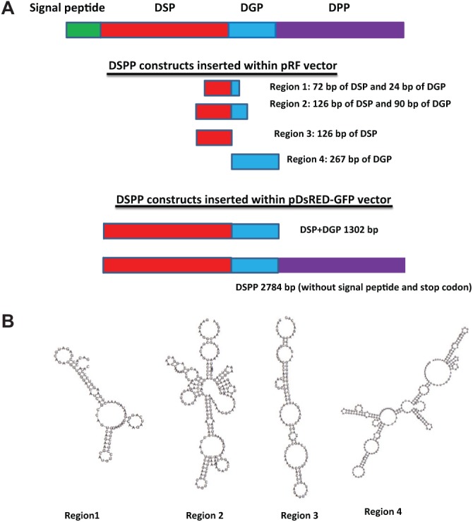
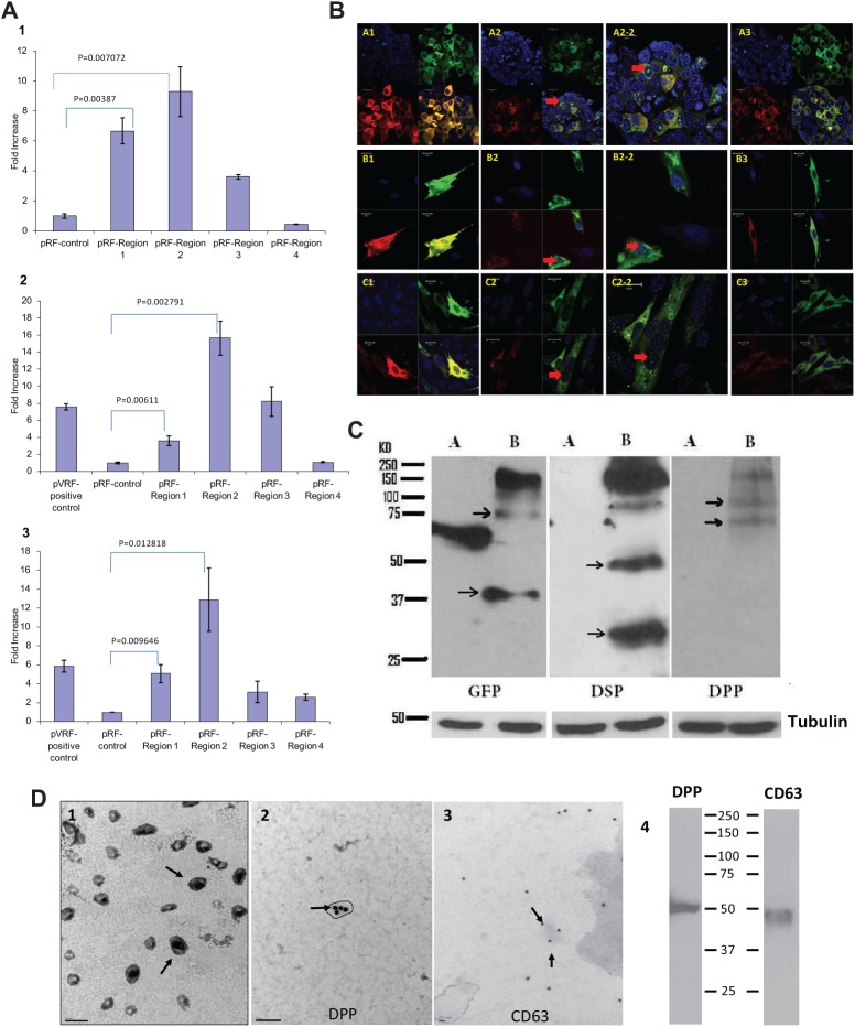
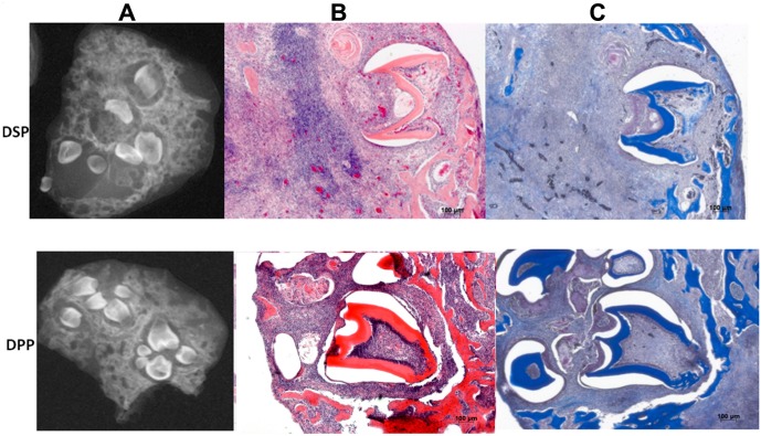
The major phosphoprotein in dentin is the aspartic acid and serine-rich protein called dentin phosphophoryn (DPP). DPP appears to be synthesized as a part of a larger compound protein, dentin sialophosphoprotein (DSPP). DSPP has never been isolated or detected in dentin extracts. It is now evident that DSPP is a chimeric protein composed of 3 parts: dentin sialoprotein (DSP), DPP, and dentin glycoprotein (DGP). Previous reports have suggested that the BMP1 protease is responsible for processing DSPP. However, unequal amounts of these products are present in the dentin matrix. Here, we provide evidence for an internal ribosome entry site in the DSPP gene that directs the synthesis of DPP. This mechanism would account for unequal amounts of intracellular DSP and DPP. The internal ribosomal entry site (IRES) activity varied in different cell types, suggesting the presence of additional regulatory elements during the translational regulation of DPP. Further, we provide evidence that DPP is transported to the extracellular matrix (ECM) through exosomes. Using tissue recombination and lentivirus-mediated gain-of-function approaches, we also demonstrate that DPP is essential for the formation of well-defined tooth structures with mineralized dentin matrix.
Keywords: cell biology; cell differentiation; dentin; extracellular matrix (ECM); matrix biology; odontoblast(s).
Conflict of interest statement
The authors declare no potential conflicts of interest with respect to the authorship and/or publications of this article.
Figures

Similar articles
-
Dentin sialophosphoprotein: a regulatory protein for dental pulp stem cell identity and fate.Stem Cells Dev. 2014 Dec 1;23(23):2883-94. doi: 10.1089/scd.2014.0066. Epub 2014 Aug 21. Stem Cells Dev. 2014. PMID: 25027178 Free PMC article.
-
Role of the NH2 -terminal fragment of dentin sialophosphoprotein in dentinogenesis.Eur J Oral Sci. 2013 Apr;121(2):76-85. doi: 10.1111/eos.12020. Epub 2013 Feb 7. Eur J Oral Sci. 2013. PMID: 23489896 Free PMC article.
-
Nuclear factor I-C (NFIC) regulates dentin sialophosphoprotein (DSPP) and E-cadherin via control of Krüppel-like factor 4 (KLF4) during dentinogenesis.J Biol Chem. 2014 Oct 10;289(41):28225-36. doi: 10.1074/jbc.M114.568691. Epub 2014 Aug 19. J Biol Chem. 2014. PMID: 25138274 Free PMC article.
-
Dentin sialophosphoprotein in biomineralization.Connect Tissue Res. 2010 Oct;51(5):404-17. doi: 10.3109/03008200903329789. Connect Tissue Res. 2010. PMID: 20367116 Free PMC article. Review.
-
Isolated dentinogenesis imperfecta and dentin dysplasia: revision of the classification.Eur J Hum Genet. 2015 Apr;23(4):445-51. doi: 10.1038/ejhg.2014.159. Epub 2014 Aug 13. Eur J Hum Genet. 2015. PMID: 25118030 Free PMC article. Review.
Cited by
-
New frontiers of oral sciences: Focus on the source and biomedical application of extracellular vesicles.Front Bioeng Biotechnol. 2022 Oct 19;10:1023700. doi: 10.3389/fbioe.2022.1023700. eCollection 2022. Front Bioeng Biotechnol. 2022. PMID: 36338125 Free PMC article. Review.
-
Survey of dentin sialophosphoprotein and its cognate matrix metalloproteinase-20 in human cancers.Cancer Med. 2019 May;8(5):2167-2178. doi: 10.1002/cam4.2117. Epub 2019 Apr 1. Cancer Med. 2019. PMID: 30932369 Free PMC article.
-
DPP an extracellular matrix molecule induces Wnt5a mediated signaling to promote the differentiation of adult stem cells into odontogenic lineage.Sci Rep. 2024 Oct 31;14(1):26187. doi: 10.1038/s41598-024-76069-7. Sci Rep. 2024. PMID: 39478025 Free PMC article.
-
DPP promotes odontogenic differentiation of DPSCs through NF-κB signaling.Sci Rep. 2021 Nov 11;11(1):22076. doi: 10.1038/s41598-021-01359-3. Sci Rep. 2021. PMID: 34764323 Free PMC article.
-
Exosomes in Extracellular Matrix Bone Biology.Curr Osteoporos Rep. 2018 Feb;16(1):58-64. doi: 10.1007/s11914-018-0419-y. Curr Osteoporos Rep. 2018. PMID: 29372401 Free PMC article. Review.
References
-
- Alvares K, Kanwar YS, Veis A. (2006). Expression and potential role of dentin phosphophoryn (DPP) in mouse embryonic tissues involved in epithelial-mesenchymal interactions and branching morphogenesis. Dev Dyn 235:2980-2990. - PubMed
-
- Bailleul-Forestier I, Molla M, Verloes A, Berdal A. (2008). The genetic basis of inherited anomalies of the teeth. Part 1: Clinical and molecular aspects of non-syndromic dental disorders. Eur J Med Genet 51:273-291. - PubMed
-
- Bègue-Kirn C, Krebsbach PH, Bartlett JD, Butler WT. (1998). Dentin sialoprotein, dentin phosphoprotein, enamelysin and ameloblastin: tooth-specific molecules that are distinctly expressed during murine dental differentiation. Eur J Oral Sci 106:963-970. - PubMed
Publication types
MeSH terms
Substances
Grants and funding
LinkOut - more resources
Full Text Sources
Other Literature Sources
Miscellaneous
