Review
. 2013 Dec 18;3(12):130181.
doi: 10.1098/rsob.130181.

Neuron-glia crosstalk in health and disease: fractalkine and CX3CR1 take centre stage

Affiliations
Review

Neuron-glia crosstalk in health and disease: fractalkine and CX3CR1 take centre stage

Graham K Sheridan et al. Open Biol. .

Abstract

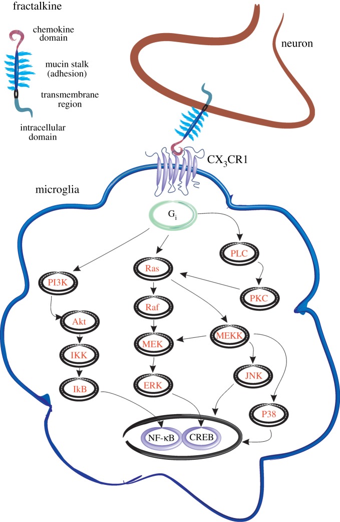
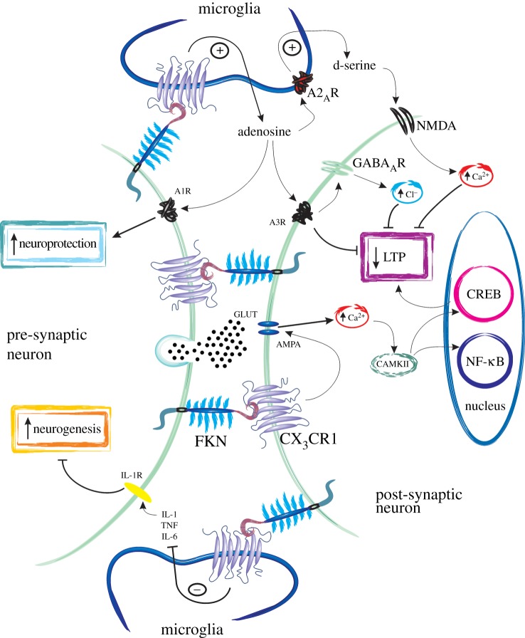
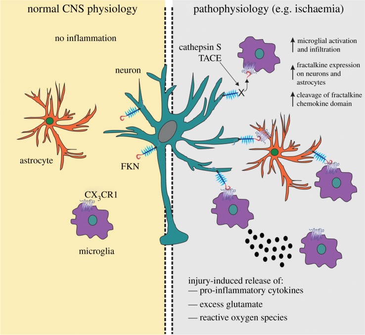
An essential aspect of normal brain function is the bidirectional interaction and communication between neurons and neighbouring glial cells. To this end, the brain has evolved ligand-receptor partnerships that facilitate crosstalk between different cell types. The chemokine, fractalkine (FKN), is expressed on neuronal cells, and its receptor, CX(3)CR1, is predominantly expressed on microglia. This review focuses on several important functional roles for FKN/CX(3)CR1 in both health and disease of the central nervous system. It has been posited that FKN is involved in microglial infiltration of the brain during development. Microglia, in turn, are implicated in the developmental synaptic pruning that occurs during brain maturation. The abundance of FKN on mature hippocampal neurons suggests a homeostatic non-inflammatory role in mechanisms of learning and memory. There is substantial evidence describing a role for FKN in hippocampal synaptic plasticity. FKN, on the one hand, appears to prevent excess microglial activation in the absence of injury while promoting activation of microglia and astrocytes during inflammatory episodes. Thus, FKN appears to be neuroprotective in some settings, whereas it contributes to neuronal damage in others. Many progressive neuroinflammatory disorders that are associated with increased microglial activation, such as Alzheimer's disease, show disruption of the FKN/CX(3)CR1 communication system. Thus, targeting CX(3)CR1 receptor hyperactivation with specific antagonists in such neuroinflammatory conditions may eventually lead to novel neurotherapeutics.

Keywords: Alzheimer's disease; CX3CR1; fractalkine; ischaemia; microglia; synaptic plasticity.


Figures

Figure 1.
Figure 1.
Fractalkine and CX3CR1 expression and signalling. Fractalkine is a large chemokine molecule consisting of four major functional regions. These include an N-terminal chemokine domain which can be cleaved by metalloproteinases such as ADAM10, TACE and the lysosomal cysteine protease, cathepsin S. The glycosylated mucin-like stalk is thought to be involved in cell adhesion, with particular affinity for migrating leucocytes at sites of inflammation. Fractalkine also contains a hydrophobic transmembrane region and intracellular C-terminal domain. Neuronally expressed membrane-bound and soluble cleaved fractalkine can bind to its receptor, CX3CR1, which is G protein-coupled and transduces several well-characterized signalling pathways leading to activation of transcription factors, including NF-κB and CREB. FKN, fractalkine; Gi, heterotrimeric G protein-coupled to Gi protein; PI3K, phosphatidylinositide 3-kinases; Ras, small GTPase; Raf, small GTPase; PLC, phospholipase C; PKC, protein kinase C; Akt, serine/threonine-specific protein kinase; MEK, mitogen-activated protein kinase kinase; MEKK, MAP3kinase; p38, p38 mitogen-activated protein kinase; IkB, inhibitor of kappa B; IKK, inhibitor of kappa B kinase; ERK, extracellular signal-regulated kinases; JNK, c-Jun N-terminal kinase; NFκB, nuclear factor kappa B; CREB, cAMP response element-binding protein.
Figure 2.
Figure 2.
Fractalkine and CX3CR1 in synaptic plasticity, neurogenesis and neuroprotection. Schematic diagram describing several mechanisms of action of fractalkine (FKN) in modulating neuronal function. Hippocampal neurons, in particular, express high levels of FKN and CX3CR1 receptors. Microglia also possess CX3CR1 and can release several chemicals that modulate neurotransmission and synaptic plasticity. First, FKN acting through CX3CR1 modulates AMPA receptor phosphorylation leading to increased calcium (Ca2+) entry and inhibition of both excitatory post-synaptic potentials (EPSPs) and long-term potentiation (LTP). FKN can also increase inhibitory post-synaptic currents (IPSCs), possibly by enhancing neuronal responsiveness to GABA-mediated chloride entry. How FKN enhances IPSCs remains unknown, but this may be due to FKN activating CX3CR1 on microglia and causing the release of adenosine. This, in turn, could activate A3R receptors on neurons, kick-starting a signalling cascade which results in modulation of GABAA receptors to increase their sensitivity to GABA. Adenosine may also activate A2AR on microglial cells and induce the release of d-serine which acts as a co-agonist at the NMDA receptor leading to increased calcium entry. In this way, FKN may also inhibit LTP induction and modulate synaptic plasticity. The adenosine released by microglia has also been suggested to play a role in neuroprotection by activating A1R receptor subtypes on neurons. Finally, FKN may play a role in hippocampal neurogenesis by inhibiting the release of IL-1β from microglial cell types. Much of this schematic diagram is speculative and based on our limited current knowledge of the interplay between FKN and CX3CR1 in CNS neurotransmission. There is still much work to be done to dissect the signalling cascades involved in FKN-mediated neuromodulation.
Figure 3.
Figure 3.
Fractalkine and CX3CR1 in neuroinflammatory conditions. In the uninjured brain under normal physiological conditions, fractalkine (FKN) is largely expressed on neurons and CX3CR1 receptors on microglial cells. FKN sequesters microglia in a quiescent ‘inactive’ state. Astrocytes are relatively devoid of FKN and CX3CR1 protein expression. Under a pathological insult, such as occurs following ischaemia, FKN can be upregulated on neuronal cells. FKN can also be cleaved by the metalloproteinase, TNFα-converting enzyme (TACE), and lysosomal cysteine protease, cathepsin S, released during injury. Upregulated levels of FKN can attract microglia to the site of inflammation, where they become activated and release pro-inflammatory mediators such as cytokines, reactive oxygen species (ROS) and glutamate. Astrocytes can also express FKN following an inflammatory insult and thus can communicate with both neurons and microglia via CX3CR1. The increased expression of FKN should have a net anti-inflammatory action and serve to limit inflammation in favour of functional recovery of CNS tissue.

References

    1. Kriegstein AR, Götz M. 2003. Radial glia diversity: a matter of cell fate. Glia 43, 37–43 (doi:10.1002/glia.10250) - DOI - PubMed
    1. Paukert M, Bergles DE. 2006. Synaptic communication between neurons and NG2+ cells. Curr. Opin. Neurobiol. 16, 515–521 (doi:10.1016/j.conb.2006.08.009) - DOI - PubMed
    1. Nishiyama A. 2007. Polydendrocytes: NG2 cells with many roles in development and repair of the CNS. Neuroscientist 13, 62–76 (doi:10.1177/1073858406295586) - DOI - PubMed
    1. Pinto L, Götz M. 2007. Radial glial cell heterogeneity: the source of diverse progeny in the CNS. Prog. Neurobiol. 83, 2–23 (doi:10.1016/j.pneurobio.2007.02.010) - DOI - PubMed
    1. Kettenmann H, Verkhratsky A. 2008. Neuroglia: the 150 years after. Trends Neurosci. 31, 653–659 (doi:10.1016/j.tins.2008.09.003) - DOI - PubMed

LinkOut - more resources