. 2014 May 1;23(9):2447-58.
doi: 10.1093/hmg/ddt640. Epub 2013 Dec 18.

Mice with an isoform-ablating Mecp2 exon 1 mutation recapitulate the neurologic deficits of Rett syndrome

Affiliations

Mice with an isoform-ablating Mecp2 exon 1 mutation recapitulate the neurologic deficits of Rett syndrome

Dag H Yasui et al. Hum Mol Genet. .

Erratum in

  • Hum Mol Genet. 2014 Dec 15;23(24):6695

Abstract

Mutations in MECP2 cause the neurodevelopmental disorder Rett syndrome (RTT OMIM 312750). Alternative inclusion of MECP2/Mecp2 exon 1 with exons 3 and 4 encodes MeCP2-e1 or MeCP2-e2 protein isoforms with unique amino termini. While most MECP2 mutations are located in exons 3 and 4 thus affecting both isoforms, MECP2 exon 1 mutations but not exon 2 mutations have been identified in RTT patients, suggesting that MeCP2-e1 deficiency is sufficient to cause RTT. As expected, genetic deletion of Mecp2 exons 3 and/or 4 recapitulates RTT-like neurologic defects in mice. However, Mecp2 exon 2 knockout mice have normal neurologic function. Here, a naturally occurring MECP2 exon 1 mutation is recapitulated in a mouse model by genetic engineering. A point mutation in the translational start codon of Mecp2 exon 1, transmitted through the germline, ablates MeCP2-e1 translation while preserving MeCP2-e2 production in mouse brain. The resulting MeCP2-e1 deficient mice developed forelimb stereotypy, hindlimb clasping, excessive grooming and hypo-activity prior to death between 7 and 31 weeks. MeCP2-e1 deficient mice also exhibited abnormal anxiety, sociability and ambulation. Despite MeCP2-e1 and MeCP2-e2 sharing, 96% amino acid identity, differences were identified. A fraction of phosphorylated MeCP2-e1 differed from the bulk of MeCP2 in subnuclear localization and co-factor interaction. Furthermore, MeCP2-e1 exhibited enhanced stability compared with MeCP2-e2 in neurons. Therefore, MeCP2-e1 deficient mice implicate MeCP2-e1 as the sole contributor to RTT with non-redundant functions.


Figures

Figure 1.
Figure 1.
Targeting strategy for MeCP2-e1-specific germline ablation. Site directed mutagenesis was used to alter the exon 1 translational start site ATG to TTG in the Mecp2 locus excised from a genomic BAC clone. A neomycin resistance gene (NEO) flanked by flippase recognition target sites (FRT) was inserted into exon 1 to produce the targeting vector (top) which was then electroporated in C57BL/6N ES cells for homologous recombination. ES cells incorporating the targeting vector were selected for neomycin resistance then the NEO cassette was excised by expressing flippase, producing the mutant allele (middle). PCR primers detecting the mutant and WT Mecp2 alleles are shown as colored arrows. Genotyping PCR was performed from DNA obtained from tail snips.
Figure 2.
Figure 2.
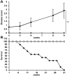
Mice with MeCP2-e1 isoform ablation exhibit neurologic disease and reduced lifespan. (A). Male Mecp2-e1−/y mice (n = 8) (black line) were scored along with control Mecp2-e1+/y male littermates (n = 6) (gray line) at weekly intervals for disease symptoms including matted fur, reduced ambulation, open skin sores, abnormally large abdomen along with hindlimb clasping, forelimb ‘washing’ and lack of body curling in response to tail suspension. Thus, the most diseased mice score ‘7’ while completely healthy mice score ‘0’. Error bars correspond to standard error of the mean (SEM). *P = 0.0197 by independent sample, two-tailed t-test. (B). Survival curve of Mecp2-e1−/y MeCP2-e1 deficient (black line) and WT control male littermate mice (gray line). n = 11 for Mecp2-e1−/y males and n = 9 for WT Mecp2-e1+/y control males.
Figure 3.
Figure 3.
Mecp2-e1−/y mice exhibit behavioral abnormalities. (A). Anxiety measured in the elevated and maze (EPM) showed increased time in the open arms and thus reduced anxiety in Mecp2-e1−/y male mice (P = 0.03*). (B) Sociability measured in the three-chamber assay revealed a significant reduction in the percent time Mecp2-e1−/y mice spent near a cage containing a mouse relative to the total time spent near an occupied or an empty cage (P = 0.048*). (C). TreadScan analysis using a transparent treadmill belt combined with a high-speed video capture to measure instantaneous running speed and foot placement found that Mecp2-e1−/y male mice were significantly slower compared with WT controls (P = 0.03*). (D). TreadScan analysis revealed significantly reduced standard deviation in the rear track width distance between rear foot stance in Mecp2-e1−/y mice compared with WT controls (P = 0.042*). Error bars correspond to SEM. Genotype group comparisons were performed using least squares ANOVA. n = 7 Mecp2-e1−/y male mice, n = 5 WT MeCP2-e1+/y male littermate controls.
Figure 4.
Figure 4.
MeCP2-e1 is ablated in Mecp2-e1−/y mouse brain, while MeCP2-e2 levels are elevated as shown by IF. (A). IF detection of MeCP2-e1 in mouse hippocampus using a MeCP2-e1 JL specific antibody in adult Mecp2-e1−/y mutant and Mecp2-e1+/y control brain sections. Alexa 594-tagged (red) secondary antibodies were used to visualize the distribution of MeCP2-e1 in the nucleus. Higher magnification images are shown in Supplementary Material, Figure S1. (B). IF detection of MeCP2-e2 in mouse hippocampus using an MeCP2-e2 antibody with Alexa 488 (green) conjugated secondary in Mecp2-e1−/y and WT Mecp2-e1+/y control hippocampal sections. DAPI staining as shown in blue reveals the location of the nucleus in all panels. The scale bar represents 5 microns.
Figure 5.
Figure 5.
Western blot analysis of brain MeCP2 isoform levels confirms loss of MeCP2-e1 and overexpression of MeCP2-e2. Total brain proteins from Mecp2-e1−/y and Mecp2-e1+/y control males were probed with an MeCP2-e1 CS specific antibody in (A) which shows barely detectable MeCP2-e1 levels in Mecp2-e1−/y brain and significantly higher levels in WT control male brain (P = 0.0019). (B) An anti-MeCP2-e2-specific antibody revealed significant elevation of this isoform (P = 0.0005) in Mecp2-e1−/y brain compared with WT (Mecp2-e1+/y) controls. (C) To determine the combined levels of MeCP2-e1 and MeCP2-e2 isoforms, an antibody recognizing a common epitope (Fig. S1) revealed an ∼2-fold reduction of total MeCP2 in Mecp2-e1−/y deficient brain by western analysis (P = 0.01). Bar graphs depict average signal intensity from NIH Image J analysis of western blots with error bars corresponding to SEM. Three to four individual mouse brain protein extracts per genotype were analyzed.
Figure 6.
Figure 6.
MeCP2-e1-associated protein identification by mass spectrometric analysis. (A) SDS–PAGE resolved MeCP2 protein complexes were purified from human SH-SY5Y neurons using MeCP2-e1 JL antibody. Proteins not co-migrating with IP antibody heavy and light chains (red boxes) were selected for proteomic analysis. A control antibody IP with pre-immune IgY is shown on the right. Molecular weight standards are shown for estimation of molecular size in kilo Daltons. (B) Selected MeCP2-e1 associated factors identified by mass spectrometric analysis. Factors were listed based on significant amino acids identified (sequence coverage) in two separate experiments (replicate). YB-1, which was previously found to be associated with MeCP2, is shown as a control.
Figure 7.
Figure 7.
MeCP2-e1 co-localizes with multiple nuclear matrix factors. (A) IF staining in cortical neurons for Matrin 3 (red) shows co-localization with MeCP2-e1 (green) distribution as shown by overlapping signal (yellow) in the merge panel. (B) Co-staining for SFPQ (red) and MeCP2-e1 (green) similarly shows significant signal overlap (yellow) indicative of co-localization in neuronal nuclei. (C). As a positive control, a previously identified MeCP2-associated factor YB-1 (red) also associates with the nuclear matrix as represented by SFPQ signal (green). Overlap of signal distribution is shown in the merged image. In all panels, the location of the nucleus is shown by DAPI counter staining (blue). Scale bars correspond to 5 μm.
Figure 8.
Figure 8.
MeCP2-e1 is more stable than MeCP2-e2 in neurons. (A) Western blot analysis of flag-tagged MeCP2-e1 and MeCP2-e2 isoform abundance following shutdown of protein expression with doxycycline treatment shown as hours (HRS). Western blot analysis of GAPDH is shown below for normalization. (B) The ratio of MeCP2-e1 (black line) and MeCP2-e2 (gray line) compared with GAPDH remaining after expression shutdown is graphed with error bars corresponding to standard error of the mean (SEM).
Figure 9.
Figure 9.
A model of MeCP2-e1 function in neurons. In this model, non-pS30 MeCP2-e1 forms a molecular complex with RNA regulatory factors on the nuclear matrix in the neuronal nucleoplasm. Neuronal signals trigger phosphorylation of MeCP2 at phosphoserine 30 leading to dissociation from the nuclear matrix and relocalization of MeCP2 to heterochromatin in chromocenters.

Similar articles

Cited by

References

    1. Amir R.E., Van den Veyver I.B., Wan M., Tran C.Q., Francke U., Zoghbi H.Y. Rett syndrome is caused by mutations in X-linked MECP2, encoding methyl-CpG-binding protein 2. Nat. Genet. 1999;23:185–188. - PubMed
    1. Coy J.F., Sedlacek Z., Bachner D., Delius H., Poustka A. A complex pattern of evolutionary conservation and alternative polyadenylation within the long 3″-untranslated region of the methyl-CpG-binding protein 2 gene (MeCP2) suggests a regulatory role in gene expression. Hum. Mol. Genet. 1999;8:1253–1262. - PubMed
    1. Kriaucionis S., Bird A. The major form of MeCP2 has a novel N-terminus generated by alternative splicing. Nucleic Acids Res. 2004;32:1818–1823. - PMC - PubMed
    1. Mnatzakanian G.N., Lohi H., Munteanu I., Alfred S.E., Yamada T., MacLeod P.J., Jones J.R., Scherer S.W., Schanen N.C., Friez M.J., et al. A previously unidentified MECP2 open reading frame defines a new protein isoform relevant to Rett syndrome. Nat. Genet. 2004;36:339–341. - PubMed
    1. Zachariah R.M., Olson C.O., Ezeonwuka C., Rastegar M. Novel MeCP2 isoform-specific antibody reveals the endogenous MeCP2E1 expression in murine brain, primary neurons and astrocytes. PloS ONE. 2012;7:e49763. - PMC - PubMed

Publication types

Substances