. 2014 Jan;141(2):346-54.
doi: 10.1242/dev.100271. Epub 2013 Dec 18.

Notch signaling is required for the formation of mesangial cells from a stromal mesenchyme precursor during kidney development

Affiliations

Notch signaling is required for the formation of mesangial cells from a stromal mesenchyme precursor during kidney development

Scott C Boyle et al. Development. 2014 Jan.

Abstract

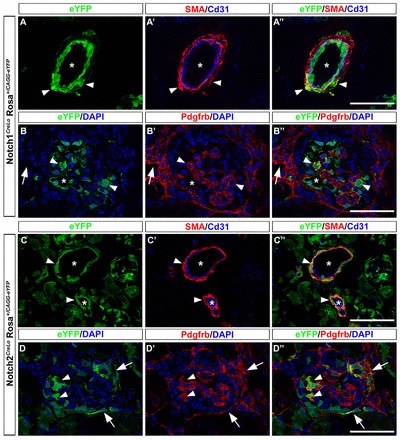
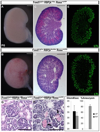
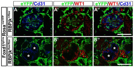
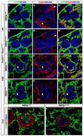
Mesangial cells are specialized pericyte/smooth muscle cells that surround and constrain the vascular network within the glomerulus of the kidney. They are derived from the stromal mesenchyme, a progenitor population distinct from nephron stem cells. Whether mesangial cells have a distinct origin from vascular smooth muscle cells (VSMCs) and the pathways that govern their specification are unknown. Here we show that Notch signaling in stromal progenitors is essential for mesangial cell formation but is dispensable for the smooth muscle and interstitial cell lineages. Deletion of RBPjk, the common DNA-binding partner of all active Notch receptors, with Foxd1(tgCre) results in glomerular aneurysm and perinatal death from kidney failure. This defect occurs early in glomerular development as stromal-derived, desmin-positive cells fail to coalesce near forming nephrons and thus do not invade the vascular cleft of the S-shaped body. This is in contrast to other mutants in which the loss of the mesangium was due to migration defects, and suggests that loss of Notch signaling results in a failure to specify this population from the stroma. Interestingly, Pdgfrb-positive VSMCs do not enter the vascular cleft and cannot rescue the mesangial deficiency. Notch1 and Notch2 act redundantly through γ-secretase and RBPjk in this process, as individual mutants have mesangial cells at birth. Together, these data demonstrate a unique origin of mesangial cells and demonstrate a novel, redundant function for Notch receptors in mesangial cell specification, proliferation or survival during kidney development.

Keywords: Foxd1; Kidney; Mesangium; Mouse; Notch; Progenitors.


Figures

Fig. 1.
Fig. 1.
Cells derived from the stromal mesenchyme experience Notch activation during development. The stromal mesenchyme gives rise to VSMCs, mesangial cells and interstitial fibroblasts. P0 kidneys carrying the Rosa+/CAG-eYFP reporter from Notch1IP-CreLO (A-B′) and Notch2IP-CreLO (C-D′) mice were used to examine lineages arising from the stroma that had experienced Notch activation (eYFP-green). (A-A′) The entire vascular endothelium (CD31, blue) is composed of cells that have experienced Notch1 activation, demonstrating the fidelity of the system. Labeled VSMCs are infrequently found in Notch1IP-CreLO kidneys (SMA-Red, arrowheads). (B-B′) Within glomeruli, mesangial cells (Pdgfrb-Red, arrowheads) are lineage positive for Notch1 activity. The majority of Notch1 lineage+ cells are endothelial cells within capillary loops (*). Interstitial fibroblasts (Pdgfrb+, outside of glomerulus, arrows) that activated Notch1 during development are not observed. (C-C′) The entire VSMC compartment is composed of cells that experienced Notch2 activation during development (SMA-Red); Notch2 cells are never found in the endothelium. (D-D′) Mesangial cells (Pdgfrb-Red, arrowheads) are also Notch2 lineage positive and interstitial fibroblasts experience Notch2 activation during development (arrows). Scale bars: 50 μm.
Fig. 2.
Fig. 2.
RBPjk is required in the stromal mesenchyme and its derivatives for proper glomerular formation. Foxd1tgCre was used to delete RBPjk from the stromal mesenchyme during kidney development. (A,B) Whole-mount images of control (A) and mutant (B) kidneys demonstrates marked blood spots visible on the surface of mutant kidneys. (C-F) H&E (C,D) and LTL (E,F) staining reveals grossly normal kidney architecture in mutants, but a reduction in proximal tubule density in the cortex. (G,H) Higher magnification of H&E staining shows aneurysm within glomeruli in mutant kidneys. (I) Quantification of glomerular number and percentage of glomeruli that have aneurysm in control and mutant kidneys. Arrowheads, normal glomeruli; arrows, hemorrhaged glomeruli. Panels A-H are representative images across multiple experiments in which the same observations were made. Control, n=8; mutant, n=12. Scale bars: 500 μm in A-D; 50 μm in G,H.
Fig. 3.
Fig. 3.
Foxd1tgCre RPBjkF/F Rosa+/eYFP glomeruli contain podocytes and severely dilated endothelial capillary loops. (A-A′) RPBjk+/F kidneys contain glomeruli with a highly organized vascular tree (CD31, blue) surrounded by podocytes (WT1, red). Stromal derived cells are found within glomeruli (YFP, green). (B-B′) RPBjkF/F glomeruli have normal podocyte distribution surrounding a completely dilated vascular endothelium with no capillary loop organization (*). Stroma-derived cells are absent from glomeruli. Scale bars: 50 μm.
Fig. 4.
Fig. 4.
Mesangial cells are absent from Foxd1tgCre RPBjkF/F Rosa+/eYFP glomeruli. Two independent mesangial markers, Pdgfrb and desmin, were used to examine glomeruli from RPBjk+/F and RPBjkF/F mice. Pdgfrb (A-B′, red) and desmin (C-D′, red) label stroma-derived (YFP, green) mesangial cells within glomeruli and interstitial fibroblasts outside glomeruli. (A-A′,C-C′) In control animals the stereotypical mesangial tree is seen and the interstitial space is filled with fibroblasts. (B-B′,D-D′) In kidneys with RBPjk deleted from stromal mesenchyme derivatives the interstitium forms normally, but the mesangium is completely absent. Occasionally, desmin+ cells were found within glomeruli, but these were invariably eYFP-, indicative of ‘escapers’ that did not express Cre (D-D′, arrowheads). Scale bars: 50 μm.
Fig. 5.
Fig. 5.
Stromal derived cells fail to enter the vascular cleft in Foxd1tgCre RPBjkF/F Rosa+/eYFP mice. (A-B′) In control kidneys cells expressing high levels of Pdgfrb (arrowheads) are found around early epithelial structures. These Pdgfrb ‘high’ cells are not evident in Foxd1tgCre RPBjkF/F Rosa+/eYFP kidneys. (C-D′) Pdgfrb+, eYFP+ stromal derived cells are absent from the vascular cleft of SSBs (arrowheads). (E,F) At the ‘cup’ stage normal podocyte patterning and vascular endothelial cells are evident in mutant kidneys, but YFP+ stromal derived cells are not found in the interior of developing glomeruli.
Fig. 6.
Fig. 6.
Desmin+ cells are absent from the area around the SSB in Foxd1tgCre RPBjkF/F Rosa+/eYFP animals. To distinguish between a mesangial migration defect (as in seen in Pdgfb/Pdgfrb mutants) and a failure of formation, we looked for desmin+ cells around developing nephrons. (A-A′) In control animals desmin+ (arrowheads) cells can be found around comma-shaped bodies and streaming into the vascular cleft. (B-B′) Desmin+ cells are absent in the area around comma-shaped bodies and from the vascular cleft in Foxd1tgCre RPBjkF/F Rosa+/eYFP kidneys.
Fig. 7.
Fig. 7.
Formation of the mesangium requires the canonical Notch signaling pathway. All Notch receptors require γ-secretase cleavage for activation. To determine if mesangial cell specification occurs through the canonical pathway or if RBPjk functions independently of Notch receptor activation we deleted PS1 and PS2, the catalytic subunits of γ-secretase. (A-A′) Mesangial cells form normally in Foxd1tgCre; PS1+/F; PS2-/-; Rosa+/R26R mice. (B-B′) Deletion of both PS1 and PS2 in Foxd1tgCre; PS1F/F; PS2-/-; Rosa+/R26R mice blocks mesangial cell development and phenocopies deletion of RPBjk. Scale bars: 50 μm.
Fig. 8.
Fig. 8.
Notch receptors act redundantly during mesangial cell formation. To determine if Notch1 or Notch2 plays a dominant role during mesangial development, we deleted each receptor individually from the SM. (A-B′) Mesangial trees in Foxd1tgCre; Notch1F/F; Rosa+/eYFP glomeruli (B-B′) are indistinguishable from control littermates (Foxd1tgCre; Notch1+/F; Rosa+/eYFP, A-A′). (C-D′) Mesangial cells form normally in Foxd1tgCre; Notch2+/F; Rosa+/eYFP (C-C′) and Foxd1tgCre; Notch2F/F; Rosa+/eYFP glomeruli (D-D′). Scale bars: 50 μm.

Similar articles

Cited by

References

    1. Alpers C. E., Hudkins K. L. (2011). Mouse models of diabetic nephropathy. Curr.Opin. Nephrol. Hypertens. 20, 278–284 - PMC - PubMed
    1. Boyle S., Misfeldt A., Chandler K. J., Deal K. K., Southard-Smith E. M., Mortlock D. P., Baldwin H. S., de Caestecker M. (2008). Fate mapping using Cited1-CreERT2 mice demonstrates that the cap mesenchyme contains self-renewing progenitor cells and gives rise exclusively to nephronic epithelia. Dev. Biol. 313, 234–245 - PMC - PubMed
    1. Boyle S. C., Kim M., Valerius M. T., McMahon A. P., Kopan R. (2011). Notch pathway activation can replace the requirement for Wnt4 and Wnt9b in mesenchymal-to-epithelial transition of nephron stem cells. Development 138, 4245–4254 - PMC - PubMed
    1. Brunskill E. W., Georgas K., Rumballe B., Little M. H., Potter S. S. (2011). Defining the molecular character of the developing and adult kidney podocyte. PLoS ONE 6, e24640 - PMC - PubMed
    1. Cheng H. T., Kim M., Valerius M. T., Surendran K., Schuster-Gossler K., Gossler A., McMahon A. P., Kopan R. (2007). Notch2, but not Notch1, is required for proximal fate acquisition in the mammalian nephron. Development 134, 801–811 - PMC - PubMed

Publication types

MeSH terms

Substances

LinkOut - more resources