. 2014 Jan;141(2):436-47.
doi: 10.1242/dev.097758. Epub 2013 Dec 18.

Postsynaptic glutamate receptors regulate local BMP signaling at the Drosophila neuromuscular junction

Affiliations

Postsynaptic glutamate receptors regulate local BMP signaling at the Drosophila neuromuscular junction

Mikolaj Sulkowski et al. Development. 2014 Jan.

Abstract

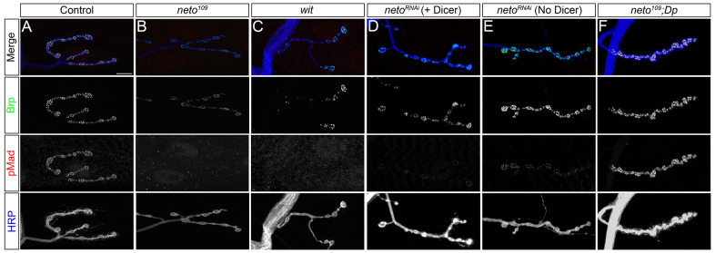
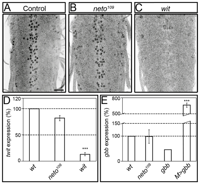
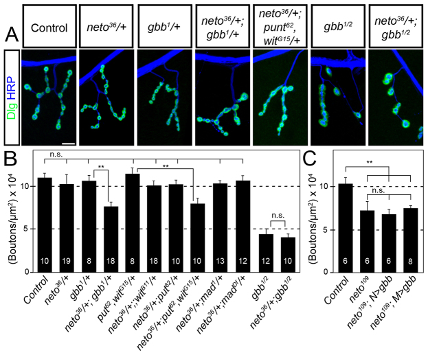
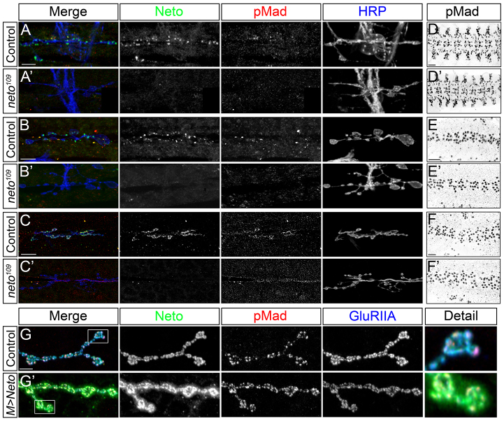
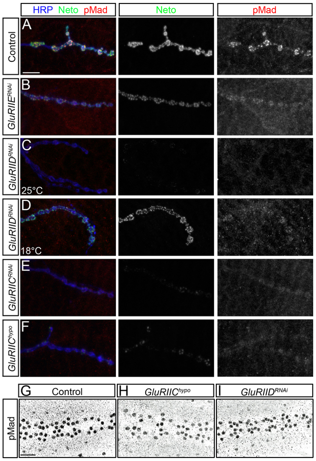
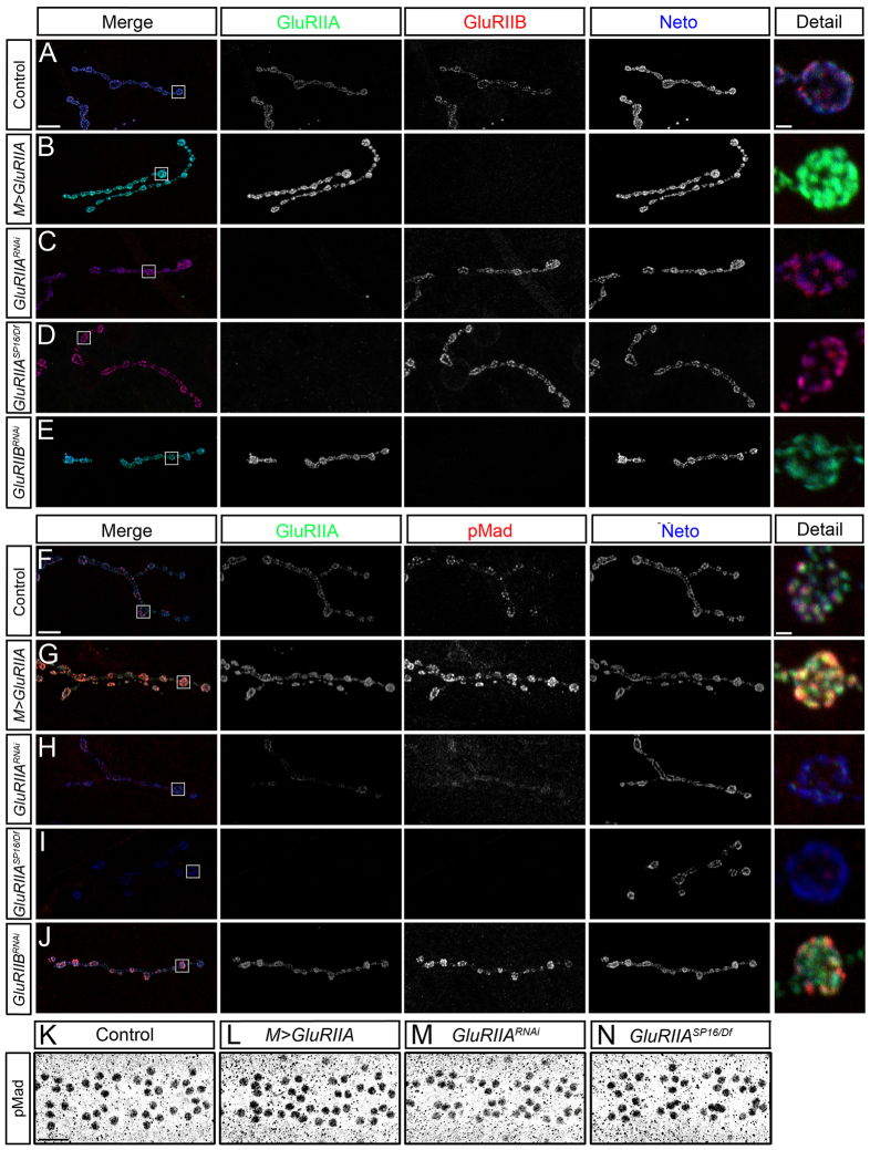
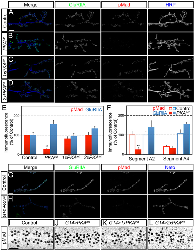
Effective communication between pre- and postsynaptic compartments is required for proper synapse development and function. At the Drosophila neuromuscular junction (NMJ), a retrograde BMP signal functions to promote synapse growth, stability and homeostasis and coordinates the growth of synaptic structures. Retrograde BMP signaling triggers accumulation of the pathway effector pMad in motoneuron nuclei and at synaptic termini. Nuclear pMad, in conjunction with transcription factors, modulates the expression of target genes and instructs synaptic growth; a role for synaptic pMad remains to be determined. Here, we report that pMad signals are selectively lost at NMJ synapses with reduced postsynaptic sensitivities. Despite this loss of synaptic pMad, nuclear pMad persisted in motoneuron nuclei, and expression of BMP target genes was unaffected, indicating a specific impairment in pMad production/maintenance at synaptic termini. During development, synaptic pMad accumulation followed the arrival and clustering of ionotropic glutamate receptors (iGluRs) at NMJ synapses. Synaptic pMad was lost at NMJ synapses developing at suboptimal levels of iGluRs and Neto, an auxiliary subunit required for functional iGluRs. Genetic manipulations of non-essential iGluR subunits revealed that synaptic pMad signals specifically correlated with the postsynaptic type-A glutamate receptors. Altering type-A receptor activities via protein kinase A (PKA) revealed that synaptic pMad depends on the activity and not the net levels of postsynaptic type-A receptors. Thus, synaptic pMad functions as a local sensor for NMJ synapse activity and has the potential to coordinate synaptic activity with a BMP retrograde signal required for synapse growth and homeostasis.

Keywords: BMP signaling; Drosophila; Glutamate receptor; Glutamatergic synapses; Neuromuscular junction.


Figures

Fig. 1.
Fig. 1.
Suboptimal Neto prevents pMad accumulation at the NMJ. (A-F) Synaptic localization of Bruchpilot (green) and pMad (red) at third instar larvae NMJ (muscle 4, abdominal segment 3). The anti-horseradish peroxidase (HRP) antibody (blue) labels motoneuron arbors. pMad localizes at NMJ synapses in control animals (A), but is diminished in neto hypomorphs, neto109 (B), similar to wit null mutants (C). Synaptic pMad signals correlate with Neto levels (D,E). A duplication containing the neto gene restores synaptic pMad at neto109 NMJs (F). Scale bars: 10 μm. Genotypes: (A) control (precise excision); (B) neto109; (C) witB11/witA12; (D) UAS-Dicer/UAS-netoRNAi-6D; 24B-Gal4/+; (E) UAS-netoRNAi-6D/+; 24B-Gal4/+; (F) neto109;; Dp(1:3)DC270.
Fig. 2.
Fig. 2.
Nuclear pMad does not change in neto hypomorphs. (A-C) Confocal images of ventral ganglia of third instar larvae immunostained against pMad. Nuclear pMad was detected in both control (A) and neto109 mutant ventral ganglia (B), including motoneuron nuclei. Nuclear pMad is lost in wit mutants (C). (D,E) Quantitative analysis of gene expression. qRT-PCR analysis reveals a small decrease of twit expression in neto109 ventral ganglia compared with controls (D). wit mutants show significantly decreased in twit expression. The levels of gbb expression in larval striated muscles are similar in neto109 and control animals, but are reduced in gbb mutants, or increased when gbb is overexpressed in the muscle (E). Bars represent the mean relative expression of target gene from three independent experiments (n=3). Error bars represent s.e.m. (***P<0.001). Genotypes: (A-D) control (precise excision), neto109, and witB11/witA12; (E) control (precise excision), neto109, gbb1/gbb2, and M>gbb (UAS-gbb9.9/G14-Gal4). Scale bar: 20 μm.
Fig. 3.
Fig. 3.
neto interacts genetically with gbb. (A) Representative confocal images of muscle 4 NMJ, abdominal segment 3, in third instar larvae immunostained against Dlg (green) and HRP (blue). Scale bar: 10 μm. (B,C) Quantification of mean number of type-IB boutons on muscle 4 per μm2 of muscle surface area. Numbers of segments analyzed are indicated. Error bars represent s.e.m. ***P<0.005 compared with control. N.S. (not significant) denotes P>0.1. Genotypes: control (precise excision), gbb1/2 (gbb1/gbb2), neto109 N>gbb (neto109; UAS-gbb9.9/+; elav-Gal4/+), neto109 M>gbb (neto109; UAS-gbb9.9/G14-Gal4).
Fig. 4.
Fig. 4.
Synaptic pMad accumulation follows Neto clustering at the developing NMJ. (A-C′) Synaptic pMad is diminished throughout development at suboptimal Neto levels. Confocal images of body wall muscles 6/7 (abdominal segment 3) using anti-Neto antibodies (green), anti-pMad (red) and anti-HRP (blue) are shown. Synaptic pMad forms puncta that colocalize with Neto puncta in late control embryos (A) but are absent in neto109 (A′). First (B) and second instar (C) larvae also show Neto and pMad synaptic signals that are largely absent from neto hypomorphs (B′,C′). (D-F′) Nuclear pMad levels remain normal at suboptimal Neto levels throughout development, including late embryos, 21 hours AEL (D,D′), first instar (E,E′) and second instar larvae (F,F′). (G,G′) High levels of Neto in muscle do not affect synaptic pMad signals. Neto labels distinct puncta that colocalize with GluRIIA signals in control animals (muscle 4, abdominal segment 3) (G). When overexpressed, Neto positive fields expand beyond the GluRIIA signals to encompass the entire bouton (G′). pMad staining does not follow the pattern of elevated Neto signals and remains colocalized with GluRIIA and Neto-positive puncta. Scale bars: 10 μm (A-C′); 20 μm (D-F′); 5 μm (G,G′). Genotypes: (G) UAS-neto-A9/+; (G′) UAS-neto-A9/G14-Gal4.
Fig. 5.
Fig. 5.
Neto/iGluR clusters control synaptic pMad accumulation. (A-F) Synaptic localization of Neto (green) and pMad (red) at third instar larvae NMJ (muscle 4, abdominal segment 3). Synaptic pMad signals are diminished when obligatory iGluR subunits are reduced via RNAi (B-E) or in hypomorphic combinations (F). Moderate reduction of synaptic Neto/iGluR complexes such as with GluRIIERNAi produce limited decrease in synaptic pMad (B). Strong loss of Neto/iGluR clusters in GluRIIDRNAi larvae induces severe reduction in the synaptic pMad signals (C); rearing GluRIIDRNAi animals at 18°C preserves some of the Neto/iGluR clusters as well as synaptic pMad (D). Reducing GluRIIC levels either by RNAi (E) or in strong hypomorphs (F) produces loss of Neto/iGluR clusters and synaptic pMad. (G-I) pMad accumulation in motoneuron nuclei remains unchanged when synaptic Neto/iGluR clusters are diminished. Nuclear pMad signals resemble controls (G) in GluRIIChypo (H) and GluRIIDRNAi (I). Scale bars: 10 μm (A-F); 20 μm (G-I). Genotypes: (A,G) control (precise excision); (B) GluRIIERNAi (G14-Gal4/+; UAS-GluRIIERNAi/+); (C,D,I) GluRIIDRNAi (G14-Gal4/+; UAS-GluRIIDRNAi/+); (E) GluRIICRNAi (G14-Gal4/+; UAS-GluRIICRNAi/+); (F,H) GluRIIEhypo (DGluRIII2/df(2L)ast1; P[UAS-cGluRIII]/+).
Fig. 6.
Fig. 6.
Type-A receptors are required for synaptic pMad accumulation. (A-J) Confocal microscopy was performed on muscle 4 (abdominal segment 3) with anti-GluRIIA antibodies (green), anti-Neto (blue) and either anti-GluRIIB (red) (A-E) or anti-pMad (red) (F-J). Overexpression of GluRIIA in striated muscles reduces GluRIIB levels compared with controls (A-B). Reduction of GluRIIA levels via RNAi (C) or in strong hypomorphs (D) increases the GluRIIB levels, whereas reduction of GluRIIB via RNAi (E) increases the GluRIIA signals. Neto synaptic signals are largely unaffected by changes in GluRIIA/GluRIIB ratio. Overexpression of GluRIIA produces an increase in pMad signal intensities compared with controls (F,G). Reducing the GluRIIA levels leads to reduction of pMad signals (H,I), but reducing the GluRIIB levels has the opposite effect (J). (K-N) Confocal microscopy of third instar ventral ganglia using anti-pMad antibodies indicates that nuclear pMad signals were not affected by manipulations of GluRIIA levels. Scale bars: 10 μm (A-J); 20 μm (K-N). Genotypes: (A,F,K) control (precise excision); (B,G,L) M>GluRIIA (UAS-GluRIIA/+; G14-Gal4/+); (C,H,M) GluRIIARNAi (G14-Gal4/+; UAS-GluRIIARNAi/+); (D,I,N) GluRIIASP16/Df (GluRIIASP16/Df(2L)clh4); (E,J) GluRIIBRNAi (G14-Gal4/+; UAS-GluRIIBRNAi/+).
Fig. 7.
Fig. 7.
GluRIIA activity controls the accumulation of synaptic pMad. (A-D) Confocal microscopy was performed on muscle 6/7 (abdominal segment 3) with anti-GluRIIA antibodies (green), anti-pMad (red) and anti-HRP (blue). Compared with control (A), muscle expression of a constitutively active catalytic subunit (PKA-act) produces a significant reduction in synaptic pMad signals (B). Expression of one (C) or two (D) copies of a mutant regulatory subunit of PKA (PKA-inh) induces a small increase in synaptic pMad accumulation. (E,F) Quantification of synaptic pMad and GluRIIA signals at various levels of muscle PKA activity. (G,H) Confocal images of third instar larvae muscle 4 (abdominal segment 3) immunostained with anti-GluRIIA antibodies (green), anti-pMad (red) and anti-Neto (blue). Strong postsynaptic PKA activity throughout development leads to complete loss of synaptic pMad and significant reduction in GluRIIA synaptic levels compared with control. (I-L) pMad accumulation in motoneuron nuclei does not change at various levels of PKA activity in the muscle. Scale bars: 10 μm (A-D); 5 μm (G,H). Genotypes: (A) control (BG487-Gal4/UAS-PKAm-inh); (B) PKAact (BG487-Gal4/UAS-PKA.mC); (C) 1xPKAinh (BG487-Gal4/+; UAS-PKAinh(GDK33)/+;); (D) 2xPKAinh (BG487-Gal4/UAS-PKAinh(GDK22); UAS-PKAinh(BDK35)/+;); (G) G14 control (G14-Gal4/UAS-PKAm-inh); (H) G14>PKAact (G14-Gal4/UAS-PKA.mC).

Similar articles

Cited by

References

    1. Aberle H., Haghighi A. P., Fetter R. D., McCabe B. D., Magalhães T. R., Goodman C. S. (2002). wishful thinking encodes a BMP type II receptor that regulates synaptic growth in Drosophila. Neuron 33, 545–558 - PubMed
    1. Adachi-Yamada T., Nakamura M., Irie K., Tomoyasu Y., Sano Y., Mori E., Goto S., Ueno N., Nishida Y., Matsumoto K. (1999). p38 mitogen-activated protein kinase can be involved in transforming growth factor beta superfamily signal transduction in Drosophila wing morphogenesis. Mol. Cell. Biol. 19, 2322–2329 - PMC - PubMed
    1. Akiyama T., Marqués G., Wharton K. A. (2012). A large bioactive BMP ligand with distinct signaling properties is produced by alternative proconvertase processing. Sci. Signal. 5, ra28 - PMC - PubMed
    1. Ball R. W., Warren-Paquin M., Tsurudome K., Liao E. H., Elazzouzi F., Cavanagh C., An B. S., Wang T. T., White J. H., Haghighi A. P. (2010). Retrograde BMP signaling controls synaptic growth at the NMJ by regulating trio expression in motor neurons. Neuron 66, 536–549 - PubMed
    1. Bellen H. J., Levis R. W., He Y., Carlson J. W., Evans-Holm M., Bae E., Kim J., Metaxakis A., Savakis C., Schulze K. L., et al. (2011). The Drosophila gene disruption project: progress using transposons with distinctive site specificities. Genetics 188, 731–743 - PMC - PubMed

Publication types

MeSH terms