. 2014 Mar;85(3):408-19.
doi: 10.1124/mol.113.090043. Epub 2013 Dec 18.

Phosphodiesterase 5 inhibitors enhance chemotherapy killing in gastrointestinal/genitourinary cancer cells

Affiliations

Phosphodiesterase 5 inhibitors enhance chemotherapy killing in gastrointestinal/genitourinary cancer cells

Laurence Booth et al. Mol Pharmacol. 2014 Mar.

Abstract

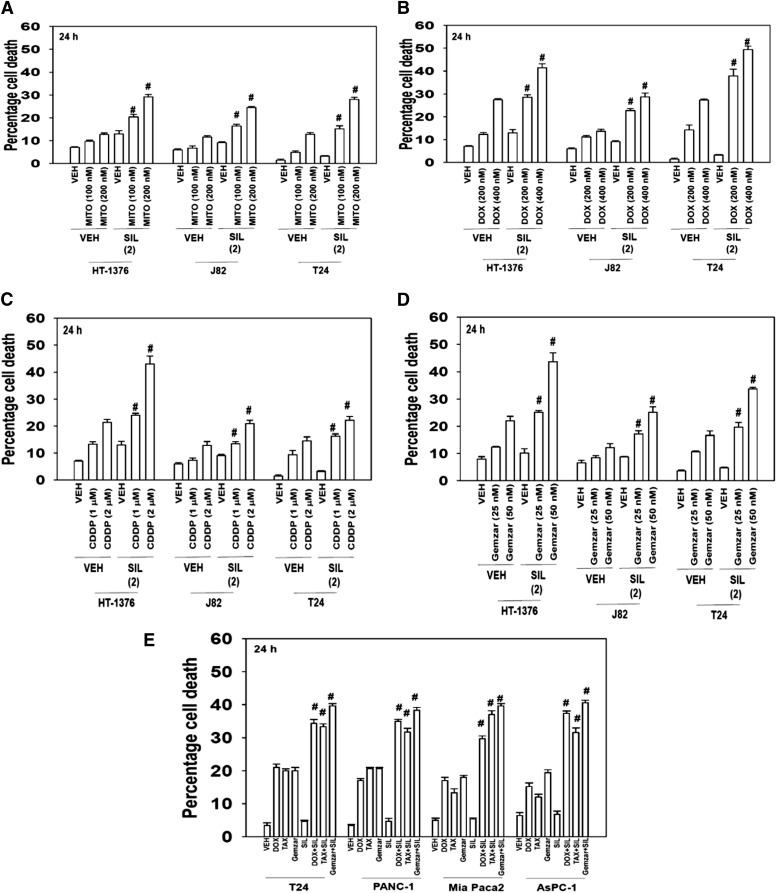
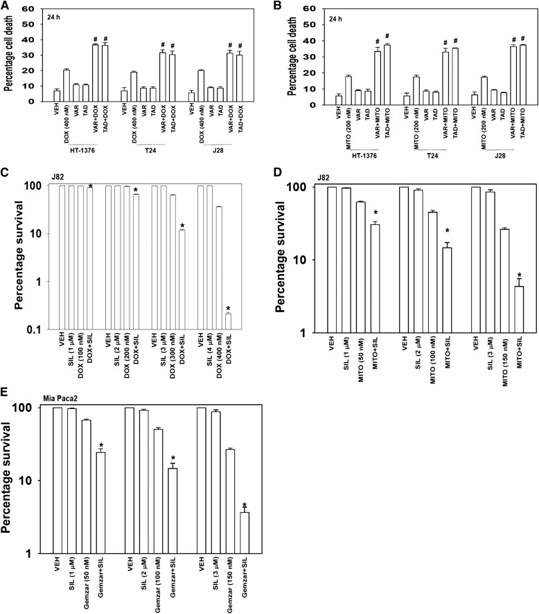
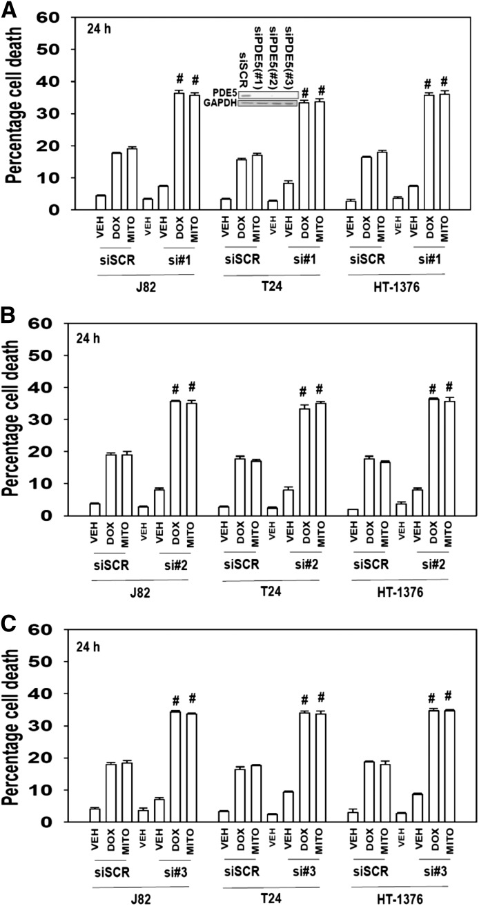
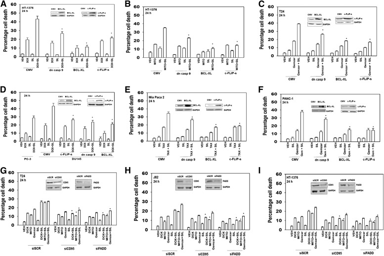
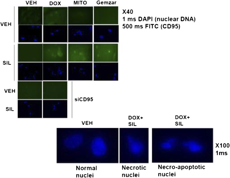
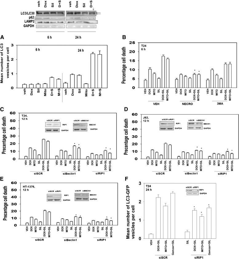
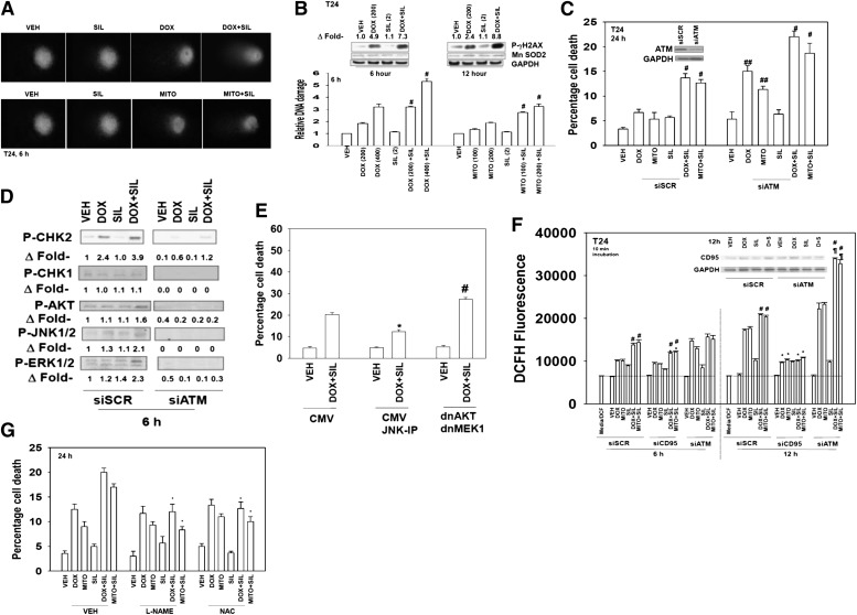
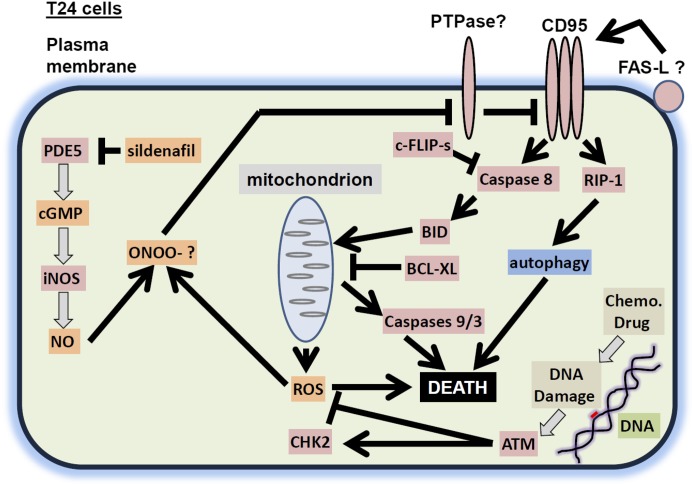
The present studies determined whether clinically relevant phosphodiesterase 5 (PDE5) inhibitors interacted with clinically relevant chemotherapies to kill gastrointestinal/genitourinary cancer cells. In bladder cancer cells, regardless of H-RAS mutational status, at clinically achievable doses, PDE5 inhibitors interacted in a greater than additive fashion with doxorubicin/mitomycin C/gemcitabine/cisplatin/paclitaxel to cause cell death. In pancreatic tumor cells expressing mutant active K-RAS, PDE5 inhibitors interacted in a greater than additive fashion with doxorubicin/gemcitabine/paclitaxel to cause cell death. The most potent PDE5 inhibitor was sildenafil. Knock down of PDE5 expression recapitulated the combination effects of PDE5 inhibitor drugs with chemotherapy drugs. Expression of cellular FLICE-like inhibitory protein-short did not significantly inhibit chemotherapy lethality but did significantly reduce enhanced killing in combination with sildenafil. Overexpression of B-cell lymphoma-extra large suppressed individual and combination drug toxicities. Knock down of CD95 or Fas-associated death domain protein suppressed drug combination toxicity. Combination toxicity was also abolished by necrostatin or receptor interacting protein 1 knock down. Treatment with PDE5 inhibitors and chemotherapy drugs promoted autophagy, which was maximal at ∼24 hour posttreatment, and 3-methyl adenine or knock down of Beclin1 suppressed drug combination lethality by ∼50%. PDE5 inhibitors enhanced and prolonged the induction of DNA damage as judged by Comet assays and γhistone 2AX (γH2AX) and checkpoint kinase 2 (CHK2) phosphorylation. Knock down of ataxia telangiectasia mutated suppressed γH2AX and CHK2 phosphorylation and enhanced drug combination lethality. Collectively our data demonstrate that the combination of PDE5 inhibitors with standard of care chemotherapy agents for gastrointestinal/genitourinary cancers represents a novel modality.


Figures

Fig. 1.
Fig. 1.
The PDE5 inhibitor sildenafil interact with established cytotoxic chemotherapy agents to kill multiple bladder cancer cell lines. (A) Bladder cancer cells (HT-1376; J82; T24) were treated with mitomycin C (MITO 100–200 nM) and/or sildenafil (SIL, 2.0 μM). Cells were isolated after 24 hours, and viability was determined by trypan blue exclusion (n = 3, ± S.E.M.). #P < 0.05 greater than corresponding value in vehicle (VEH) control. (B) Bladder cancer cells (HT-1376; J82; T24) were treated with DOX (200–400 nM) and/or SIL (2.0 μM). Cells were isolated after 24 hours, and viability was determined by trypan blue exclusion (n = 3, ± S.E.M.). #P < 0.05 greater than corresponding value in vehicle control. (C) Bladder cancer cells (HT-1376; J82; T24) were treated with cisplatin [cisplatinum (CDDP); 1000–2000 nM] and/or SIL (2.0 μM). Cells were isolated after 24 hours, and viability was determined by trypan blue exclusion (n = 3, ± S.E.M.). #P < 0.05 greater than corresponding value in vehicle control. (D) Bladder cancer cells (HT-1376; J82; T24) were treated with Gemzar (25–50 nM) and/or SIL (2.0 μM). Cells were isolated after 24 hours, and viability was determined by trypan blue exclusion (n = 3, ± S.E.M.). #P < 0.05 greater than corresponding value in vehicle control. (E) Bladder and pancreatic cancer cells (T24, PANC-1, Mia Paca2, AsPC-1) were treated with Gemzar (25 nM) and/or paclitaxel (TAX, 10 nM) and/or SIL (2.0 μM). Cells were isolated after 24 hours, and viability was determined by trypan blue exclusion (n = 3, ± S.E.M.). #P < 0.05 greater than corresponding value in vehicle control.
Fig. 2.
Fig. 2.
PDE5 inhibitors enhance doxorubicin or mitomycin C toxicity. (A) Bladder cancer cells (HT-1376; J82; T24) were treated with DOX (400 nM) and/or vardenafil (VAR, 0.5 μM) and/or tadalafil (TAD, 2 μM). Cells were isolated after 24 hours, and viability was determined by trypan blue exclusion (n = 3, ± S.E.M.). #P < 0.05 greater than corresponding value in vehicle control. (B) Bladder cancer cells (HT-1376; J82; T24) were treated with mitomycin C (MITO, 200 nM) and/or VAR (0.5 μM) and/or TAD (2 μM). Cells were isolated after 24 hours, and was viability determined by trypan blue exclusion (n = 3, ± S.E.M.). #P < 0.05 greater than corresponding value in vehicle control. (C) J82 cells were plated as single cells in sextuplicate (250–500 cells per well). Twelve hours after plating cells were treated with vehicle, sildenafil (SIL, 1–4 μM), DOX (100–400 nM), or the combination in a fixed dose ratio. Cells were treated with drugs for 24 hours followed by culture in drug-free media for ∼10 days. Colonies were fixed, stained, and counted (n = 3, ± S.E.M.). *P < 0.05 less than DOX alone value. (D) J82 cells were plated as single cells in sextuplicate (250–500 cells per well). Twelve hours after plating cells were treated with vehicle, SIL (1–3 μM), MITO (50–150 nM), or the combination in a fixed dose ratio. Cells were treated with drugs for 24 hours followed by culture in drug-free media for ∼10 days. Colonies were fixed, stained, and counted (n = 3, ± S.E.M.). *P < 0.05 less than MITO alone value. (E) Mia Paca 2 cells were plated as single cells in sextuplicate (250–500 cells per well). Twelve hours after plating cells were treated with vehicle, SIL (1–3 μM), Gemzar (50–150 nM), or the combination in a fixed dose ratio. Cells were treated with drugs for 24 hours followed by culture in drug-free media for ∼10 days. Colonies were fixed, stained, and counted (n = 3, ± S.E.M.). *P < 0.05 less than Gemzar alone value.
Fig. 3.
Fig. 3.
Knock down of PDE5 expression enhances doxorubicin or mitomycin C lethality. Bladder cancer cells (HT-1376; J82; T24) were transfected with scrambled siRNA (siSCR) or one of three different siRNA molecules to knock down expression of PDE5 (si#1, si#2, si#3; A–C). Thirty-six hours after transfection cells were treated with vehicle, mitomycin C (MITO, 100 nM), DOX (200 nM) as indicated. Cells were isolated after 24 hours, and viability was determined by trypan blue exclusion (n = 3, ± S.E.M.). #P < 0.05 greater than corresponding value in siSCR control.
Fig. 4.
Fig. 4.
The toxic interaction between PDE5 inhibitors and chemotherapy is blocked by overexpression of BCL-XL or c-FLIP-s. Bladder cancer cells (HT-1376; J82; T24) were infected with empty vector adenovirus (CMV) or three other viruses to express dominant negative caspase 9 (dn casp 9); BCL-XL; c-FLIP-s. (A–C) Thirty-six hours after infection cells were treated with vehicle, sildenafil (SIL, 2 μM), mitomycin C (MITO, 100 nM), DOX (200 nM), Gemzar (50 nM) as indicated. Cells were isolated after 24 hours, and viability was determined by trypan blue exclusion (n = 3, ± S.E.M.). *P < 0.05 less than corresponding value in CMV control. (D) Prostate cancer cells (DU145) were infected with CMV or three other viruses to express dn casp 9, BCL-XL, c-FLIP-s. Thirty-six hours after infection cells were treated with vehicle, SIL (2 μM), DOX (200 nM), as indicated. PC-3 prostate cancer cells that lack phosphatase and tensin homolog and have constitutive AKT activity were used as an internal control. Cells were isolated after 24 hours, and viability was determined by trypan blue exclusion (n = 3, ± S.E.M.). *P < 0.05 less than corresponding value in CMV control. (E and F) Pancreatic cancer cells (PANC-1, Mia Paca2) were infected with CMV or three other viruses to express dn casp 9: BCL-XL, c-FLIP-s treated with vehicle, SIL (2 μM), and/or gemcitabine (Gemzar, 50 nM) and/or paclitaxel (TAX, 10 nM) as indicated. Cells were isolated after 24 hours, and viability was determined by trypan blue exclusion (n = 3, ± S.E.M.). (G–I) Bladder cancer cells (HT-1376; J82; T24) were transfected with scrambled siRNA (siSCR) or siRNA molecules to knock down expression of CD95 or Fas-associated death domain protein (FADD) (siCD95, siFADD). Thirty-six hours after transfection cells were treated with vehicle, SIL (2 μM), MITO (100 nM), DOX (200 nM), Gemzar (50 nM) as indicated. Cells were isolated after 24 hours, and viability was determined by trypan blue exclusion (n = 3, ± S.E.M.). *P < 0.05 less than corresponding value in siSCR control.
Fig. 5.
Fig. 5.
The toxic interaction between PDE5 inhibitors and chemotherapy is associated with increased plasma membrane levels. Bladder T24 cells were grown in chamber slides. Twenty-four hours after plating cells were treated with vehicle, sildenafil (SIL, 2 μM), mitomycin C (MITO, 100 nM), DOX (200 nM), Gemzar (50 nM) as indicated. Cells were fixed 6 hours after exposure, and the cell surface levels of CD95 under each condition were determined by immunohistochemistry. DAPI, 4′,6-diamidino-2-phenylindole; FITC, fluorescein isothiocyanate.
Fig. 6.
Fig. 6.
Sildenafil and chemotherapy-induced lethality is mediated through RIP-1 and increased autophagy. (A) (Bottom) T24 cells were transfected with a plasmid to express LC3-GFP. Twenty-four hours after transfection cells were treated with vehicle, sildenafil (SIL, 2 μM), mitomycin C (MITO, 100 nM), DOX (200 nM), Gemzar (50 nM) as indicated. Cells were examined 6 and 24 hours after treatment using a fluorescent microscope, and the mean number of LC3-GFP+ vesicles was determined in >40 cells (n = 3, ± S.E.M.). (Top) T24 cells were treated with vehicle, SIL (2 μM), DOX (200 nM) as indicated. Cells were isolated 6 and 24 hours after treatment, and immunoblotting was performed to examine the phosphorylation/expression of the indicated proteins (n = 3). (B) T24 cells were treated with vehicle, SIL (2 μM), MITO (100 nM), DOX (200 nM) as indicated. In parallel, cells were treated with vehicle, necrostatin (1.0 μM) or 3-methyl adenine (3MA, 10 mM). Cells were isolated after 6 hours, and viability was determined by trypan blue exclusion (n = 3, ± S.E.M.). *P < 0.05 less than corresponding value in vehicle control. (C–E) Bladder cancer cells (HT-1376; J82; T24) were transfected with scrambled siRNA (siSCR) or siRNA molecules to knock down expression of RIP-1 or Beclin1 (siRIP-1, siBeclin1). Thirty-six hours after transfection, cells were treated with vehicle, SIL (2 μM), MITO (100 nM), or DOX (200 nM) as indicated. Cells were isolated after 12 hours, and viability was determined by trypan blue exclusion (n = 3, ± S.E.M.). *P < 0.05 less than corresponding value in siSCR control. (F) T24 cells were transfected to express LC3-GFP and with siSCR or siRNA molecules to knock down expression of RIP-1 (siRIP-1). Thirty-six hours after transfection cells were treated with vehicle, SIL (2 μM), MITO (100 nM), or DOX (200 nM) as indicated. Cells were examined 24 hours after treatment using a fluorescent microscope, and the mean number of LC3-GFP+ vesicles was determined in >40 cells (n = 3, ± S.E.M.). GAPDH, glyceraldehyde 3-phosphate dehydrogenase.
Fig. 7.
Fig. 7.
Sildenafil increases and prolongs chemotherapy-induced DNA damage; knock down of ATM enhances drug combination toxicity. (A) T24 cells were grown in soft agar and treated as indicated with vehicle, sildenafil (SIL, 2 μM), mitomycin C (MITO, 100 nM), DOX (200 nM) as indicated for 6 hours. Cells were subjected to electrophoresis and stained. Images are a representative (n = 4). (B) Graph: the tail moments of cells imaged in Awere measured and plotted (n = 4, ± S.E.M.). #P < 0.05 greater than corresponding value in vehicle-treated cells. Blot: T24 cells were treated with vehicle, SIL (2 μM), DOX (200 nM) as indicated. Cells were isolated 6 and 12 hours after exposure, and the phosphorylation of γH2AX was determined, with the fold increase in phosphorylation shown (n = 3, ± S.E.M.). (C) T24 cells were transfected with scrambled siRNA (siSCR) or siRNA molecules to knock down expression of ATM (siATM). Thirty-six hours after transfection cells were treated with vehicle, SIL (2 μM), MITO (100 nM), or DOX (200 nM) as indicated. Cells were isolated after 24 hours, and viability was determined by trypan blue exclusion (n = 3, ± S.E.M.). #P < 0.05 greater than DOX/MITO only treated cells; ##P < 0.05 greater than corresponding value in siSCR control. (D and E) Blot: T24 cells were transfected with siSCR or siRNA molecules to knock down expression of ATM (siATM). Thirty-six hours after transfection cells were treated with vehicle, SIL (2 μM), and/or DOX (200 nM) as indicated. Cells were isolated after 6 hours, and the expression and the phosphorylation of the indicated proteins was determined (n = 3). Graph: T24 cells were transfected with an empty vector plasmid (CMV) or plasmids to express dominant negative MEK1 and dominant negative AKT. Twenty-four hours after transfection, cells were as indicated treated with the JNK inhibitory peptide (JNK-IP, 10 μM) and then treated with vehicle or SIL (2 μM) and DOX (200 nM) as indicated. Cells were isolated after 24 hours, and viability was determined by trypan blue exclusion (n = 3, ± S.E.M.). #P < 0.05 greater than SIL+DOX in CMV cells; *P < 0.05 less than corresponding value in CMV. (F) T24 cells were plated in 96-well plates in phenol red free media. T24 cells were transfected with siSCR or siRNA molecules to knock down expression of ATM (siATM) or CD95 (siCD95). Thirty-six hours after transfection cells were treated with vehicle, SIL (2 μM), MITO (100 nM), or DOX (200 nM) as indicated. Cells were incubated with dihydro-dichlorofluorescein (5 mM for 10 minutes). Fluorescence measurements were obtained 6 and 12 hours after drug addition with a Vector 3 plate reader. Data are presented including basal fluorescence of vehicle-treated cells at each time point (n = 3, ± S.E.M.). #P < 0.05 greater than cells treated with only DOX/MITOs; P < 0.05 greater than corresponding value in siSCR control; *P < 0.05 less than sildenafil/doxorubicin alone. (G) Bladder cancer cells (HT-1376; J82; T24) were treated with vehicle or l-NAME (10 mM) or NAC (10 mM) followed 30 minutes later by vehicle, SIL (2 μM), MITO (100 nM), DOX (200 nM), Gemzar (50 nM) as indicated. Cells were isolated after 24 hours, and viability was determined by trypan blue exclusion (n = 3, ± S.E.M.).
Fig. 8.
Fig. 8.
A schematic of how PDE5 inhibitors and DNA damaging drugs interact to kill T24 cells. PDE5 inhibitors such as sildenafil inhibit PDE5, which leads to increased cGMP levels, increased iNOS expression, and elevated levels of NO. DNA damaging drugs stimulate the production of ROS that is counteracted by ATM. ROS and NO generate peroxynitrite, which inhibits the protein tyrosine phosphatase that inhibits CD95. CD95 becomes activated, stimulating (1) the extrinsic apoptosis pathway and (2) through RIP-1, an autophagy pathway that also leads to cell killing. BID, BH3 interacting-domain death agonist.

Similar articles

Cited by

References

    1. Amir M, Zhao E, Fontana L, Rosenberg H, Tanaka K, Gao G, Czaja MJ. (2013) Inhibition of hepatocyte autophagy increases tumor necrosis factor-dependent liver injury by promoting caspase-8 activation. Cell Death Differ 20:878–887 - PMC - PubMed
    1. Antoni L, Sodha N, Collins I, Garrett MD. (2007) CHK2 kinase: cancer susceptibility and cancer therapy - two sides of the same coin? Nat Rev Cancer 7:925–936 - PubMed
    1. Bareford MD, Park MA, Yacoub A, Hamed HA, Tang Y, Cruickshanks N, Eulitt P, Hubbard N, Tye G, Burow ME, et al. (2011) Sorafenib enhances pemetrexed cytotoxicity through an autophagy-dependent mechanism in cancer cells. Cancer Res 71:4955–4967 - PMC - PubMed
    1. Bender AT, Beavo JA. (2006) Cyclic nucleotide phosphodiesterases: molecular regulation to clinical use. Pharmacol Rev 58:488–520 - PubMed
    1. Booth L, Cruickshanks N, Ridder T, Dai Y, Grant S, Dent P. (2013) PARP and CHK inhibitors interact to cause DNA damage and cell death in mammary carcinoma cells. Cancer Biol Ther 14:458–465 - PMC - PubMed

Publication types

MeSH terms