. 2014 Mar;42(5):3177-93.
doi: 10.1093/nar/gkt1311. Epub 2013 Dec 17.

NKAP is a novel RS-related protein that interacts with RNA and RNA binding proteins

Affiliations

NKAP is a novel RS-related protein that interacts with RNA and RNA binding proteins

Bhagyashri D Burgute et al. Nucleic Acids Res. 2014 Mar.

Abstract

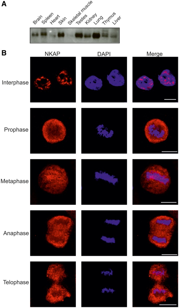
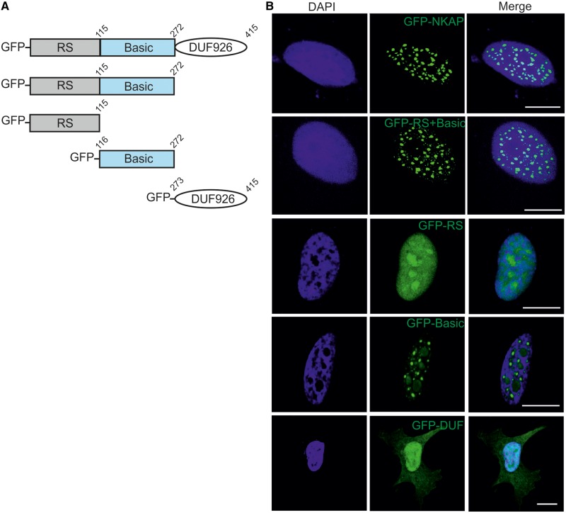
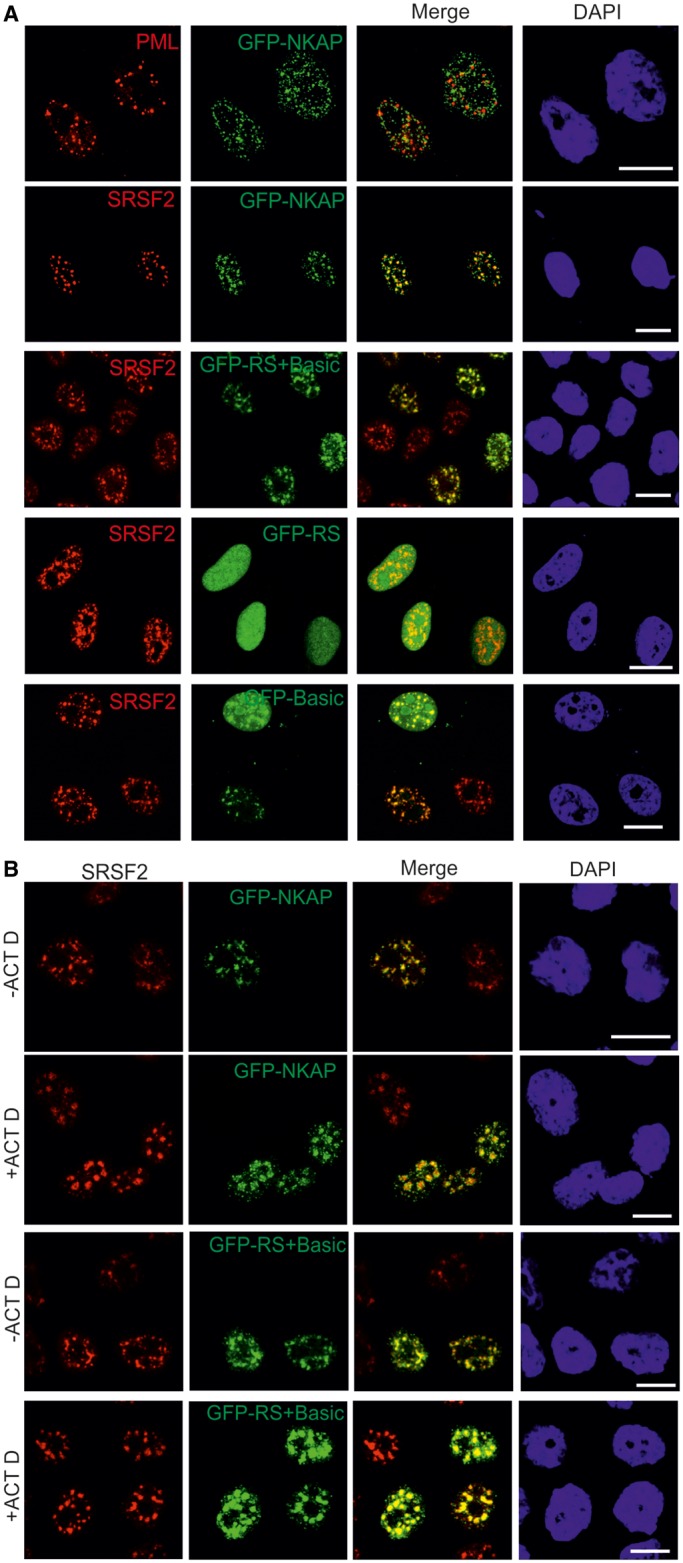
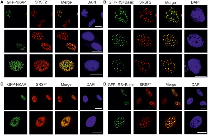
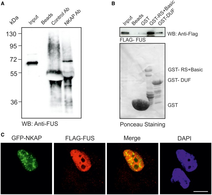
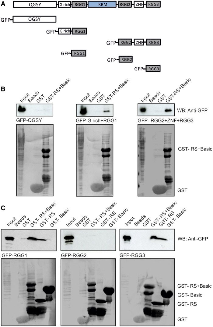
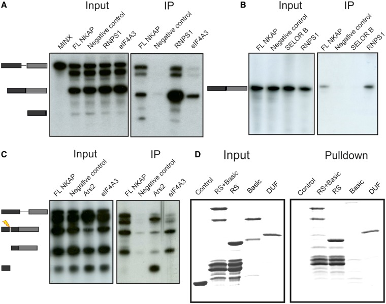
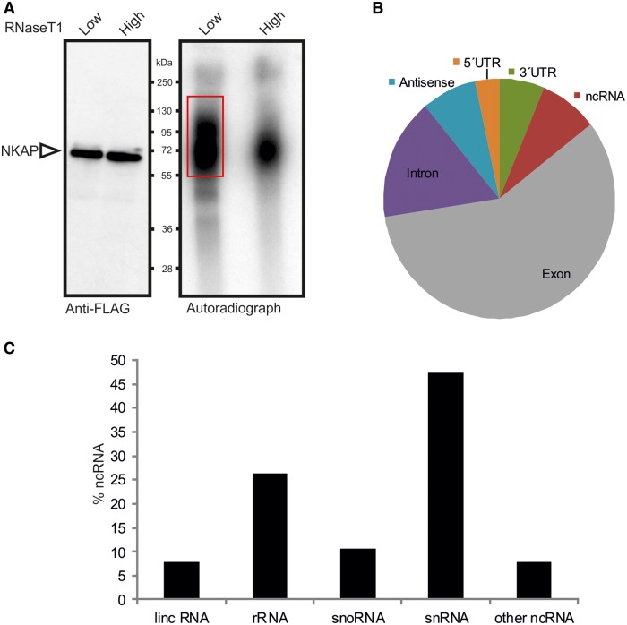
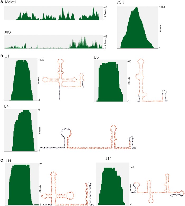
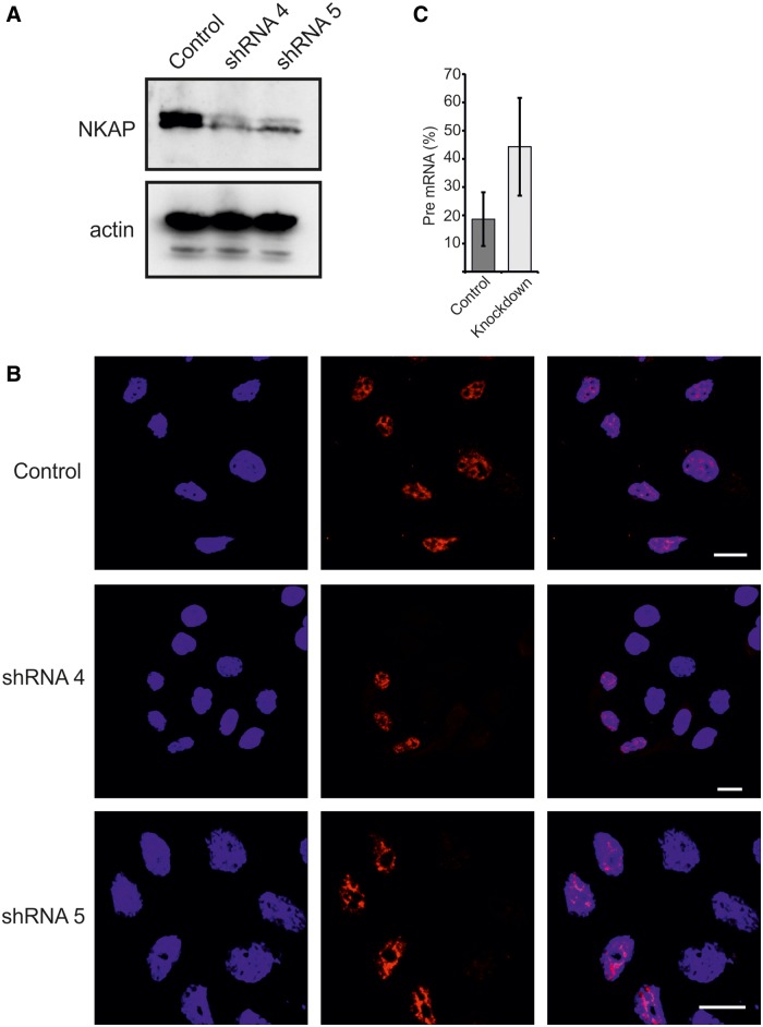
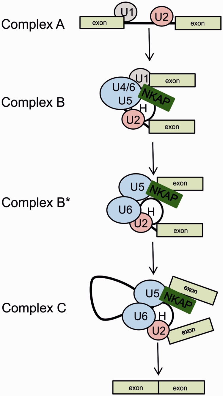
NKAP is a highly conserved protein with roles in transcriptional repression, T-cell development, maturation and acquisition of functional competency and maintenance and survival of adult hematopoietic stem cells. Here we report the novel role of NKAP in splicing. With NKAP-specific antibodies we found that NKAP localizes to nuclear speckles. NKAP has an RS motif at the N-terminus followed by a highly basic domain and a DUF 926 domain at the C-terminal region. Deletion analysis showed that the basic domain is important for speckle localization. In pull-down experiments, we identified RNA-binding proteins, RNA helicases and splicing factors as interaction partners of NKAP, among them FUS/TLS. The FUS/TLS-NKAP interaction takes place through the RS domain of NKAP and the RGG1 and RGG3 domains of FUS/TLS. We analyzed the ability of NKAP to interact with RNA using in vitro splicing assays and found that NKAP bound both spliced messenger RNA (mRNA) and unspliced pre-mRNA. Genome-wide analysis using crosslinking and immunoprecipitation-seq revealed NKAP association with U1, U4 and U5 small nuclear RNA, and we also demonstrated that knockdown of NKAP led to an increase in pre-mRNA percentage. Our results reveal NKAP as nuclear speckle protein with roles in RNA splicing and processing.


Figures

Figure 1.
Figure 1.
Expression and localization of NKAP. (A) Presence of NKAP in mouse tissues. Homogenates of adult mouse tissues as indicated were separated by SDS-PAGE (12% acrylamide) and the corresponding blot probed with NKAP-specific mAb K85-80-5. (B) Localization of NKAP during mitosis. HeLa cells were synchronized using nocodazole to block progression of the cell cycle and then released and fixed using 4% paraformaldehyde (PFA). Cells were stained with mAb K85-80-5, and nuclei (blue) were stained with 4′,6-diamidino-2-phenylindole (DAPI). The phases of the cell cycle are indicated. Bar, 10 µm.
Figure 2.
Figure 2.
Subcellular localization of NKAP. (A) Schematic representation of NKAP polypeptides used in this study. (B) Full length NKAP, Rs + Basic domain, RS domain, Basic domain and DUF 926 domain were tagged with N-terminal GFP and expressed in HeLa cells that were fixed and stained with DAPI. Bar, 10 µm.
Figure 3.
Figure 3.
NKAP localizes to the nuclear speckles. (A) HeLa cells overexpressing GFP-NKAP, GFP-RS+Basic, GFP-RS and GFP-Basic were fixed with 4% PFA and stained for PML bodies and nuclear speckles by pAB PML and mAb SRSF2, respectively. DNA was stained with DAPI. (B) Arrest of transcription affects NKAP and RS + Basic distribution. HeLa cells expressing GFP-NKAP and GFP-RS+Basic were treated with actinomycin D, fixed with 4% PFA and stained for SRSF2. Bar, 10 µm.
Figure 4.
Figure 4.
Overexpression of NKAP and RS+Basic alters SRSF2 localization. (A) HeLa cells expressing GFP-NKAP were stained for SRSF2. Cells expressing moderate amounts (upper panel) and strongly overexpressing GFP-NKAP are shown (middle and lower panel). (B) Expression of GFP-RS+Basic in HeLa cells. Upper panel, cells expressing moderate amounts, middle and lower panel, overexpression of GFP-RS+Basic. Bar, 10 µm. (C) The distribution of SRSF1 and speckle number was altered when GFP-NKAP was strongly overexpressed. (D) Overexpression of RS+Basic led to altered localization of SRSF1.
Figure 5.
Figure 5.
RS+Basic interacts with FUS. (A) Immunoprecipitation of FUS using anti-NKAP monoclonal antibody K85-80-5. (B) The GST-RS+Basic of NKAP precipitates FLAG-FUS recognized by polyclonal FLAG antibodies. GST, GST-RS+Basic and GST-DUF were visualized by Ponceau S staining. Proteins were separated by 12% SDS-PAGE. (C) HeLa cells were transfected with GFP-NKAP and FLAG-FUS, fixed and stained with FLAG antibodies. Secondary antibody conjugated with Alexa 568 was used. Nuclei were visualized with DAPI. Bar, 10 µm.
Figure 6.
Figure 6.
The RS domain of NKAP interacts with RGG1 and RGG3. (A) Schematic representation of FUS/TLS constructs used in this study. (B) GST-RS+Basic pulls down GFP-G rich+RGG1 and GFP-RGG2+ZNF+RGG3 recognized by mAb GFP K184-3. GST-RS+Basic and GST were visualized by Ponceau S staining. Proteins were separated by 12% SDS-PAGE. (C) GST-RS domain of NKAP interacts with GFP-RGG1 and GFP-RGG3. GST-RS+Basic, GST-RS, GST-Basic and GST were visualized by Ponceau S staining.
Figure 7.
Figure 7.
NKAP interacts with RNA. (A) 32P-labeled MINX pre-mRNA was spliced in vitro in HEK293T cell extracts containing FLAG-tagged proteins (RNPS1 and eIF4A3 as positive control, FLAG alone served as negative control) followed by immunoprecipitation by FLAG antibody coupled beads (IP). Aliquots of the total reactions were also analyzed (input). The positions of pre-mRNA, spliced mRNA and exon 1 (from top to bottom) are shown on the left. (B) 32P-labeled intronless mRNA MINX was incubated for 2 h in a HEK293T cell extract expressing FLAG-tagged proteins (RNPS1 as positive control, SELOR B and FLAG were negative control) followed by immunoprecipitation using FLAG antibodies coupled beads (IP). Aliquots of the total reactions were also analyzed (input). (C) 32P-labeled MINX pre-mRNA was spliced in vitro using HEK293T cell extracts expressing FLAG-tagged proteins. It was followed by addition of an oligonucleotide complementary to the first exon to activate RNase H cleavage of the RNA and to release a 5′ cap containing fragment and by immunoprecipitation with FLAG antibodies (IP). Aliquots of the total reactions were also analyzed using denaturing PAGE (Input). The mobilities of pre-mRNA, spliced mRNA, exon 1 and 5′ fragment are marked on the left. (D) RNA-binding assay using Sepharose-conjugated RNA homopolymers and GST-RS+Basic, GST-RS and GST-DUF followed by western blotting. GST was used for control.
Figure 8.
Figure 8.
Global analysis of NKAP binding to RNA by CLIP-seq. (A) Autoradiograph of cross-linked NKAP-RNA complexes using denaturing gel electrophoresis and membrane transfer. RNA was partially digested using low or high concentration of RNase T1. The marked area was cut from the membrane and subjected to protocols for the isolation of RNA, which was further used for RNA-seq. (B) Distributions of NKAP CLIP-tags. Binding regions are mapped to exons, UTRs, introns, antisense and ncRNAs according to the University of California, Santa Cruz human genome browser. Pie-charts show ratios of binding regions mapped to the indicated regions. (C) Distribution of cross-link sites within the ncRNA classes.
Figure 9.
Figure 9.
Association of NKAP with ncRNA. (A) NKAP association with Malat1, Xist and 7SK. (B and C) Interaction of NKAP with snRNAs required for splicing of both major (B) and minor (C) classes of introns. The secondary structure of each snRNA is shown on the right. The covered regions in CLIP-seq experiment of snRNAs are shown in red.
Figure 10.
Figure 10.
Role of NKAP in regulating splicing. (A) Efficient knockdown of NKAP using two different oligos. Western blotting was performed using NKAP mAB K85-80-5. (B) Control and knockdown HEK 293T cells were fixed with 4% PFA and stained using mAB K85-80-5. (C) The pre-mRNA percentage of GAPDH in control and NKAP knockdown HEK 293T cells. A significant increase was observed in knockdown cells (n = 6, P = 0.0164).
Figure 11.
Figure 11.
Assembly of spliceosomal complexes and proposed function of NKAP. Model presenting the function of NKAP and its association with snRNAs. NKAP associates with the B splicing complex, specifically with U1, U4/U5 snRNA and complex H (hnRNP proteins) and could act as a trigger to activate this complex via dissociation of U1 and U4 snRNA while keeping its tight association with U5 snRNA and complex H.

References

    1. Lamond AI, Spector DL. Nuclear speckles: a model for nuclear organelles. Nat. Rev. Mol. Cell Biol. 2003;4:605–612. - PubMed
    1. Spector DL, Lamond AI. Nuclear speckles. Cold Spring Harb. Perspect. Biol. 2011;3:a000646. - PMC - PubMed
    1. Lafarga M, Casafont I, Bengoechea R, Tapia O, Berciano MT. Cajal's contribution to the knowledge of the neuronal cell nucleus. Chromosoma. 2009;118:437–443. - PubMed
    1. Hall LL, Smith KP, Byron M, Lawrence JB. Molecular anatomy of a speckle. Anat. Rec. A Discov. Mol. Cell. Evol. Biol. 2006;288:664–675. - PMC - PubMed
    1. Melcák I, Cermanová S, Jirsová K, Koberna K, Malínský J, Raska I. Nuclear pre-mRNA compartmentalization: trafficking of released transcripts to splicing factor reservoirs. Mol. Biol. Cell. 2000;11:497–510. - PMC - PubMed

Publication types