

Automatic corpus callosum segmentation using a deformable active Fourier contour model
- PMID: 24353382
- PMCID: PMC3864934
- DOI: 10.1117/12.911504
Automatic corpus callosum segmentation using a deformable active Fourier contour model
Abstract
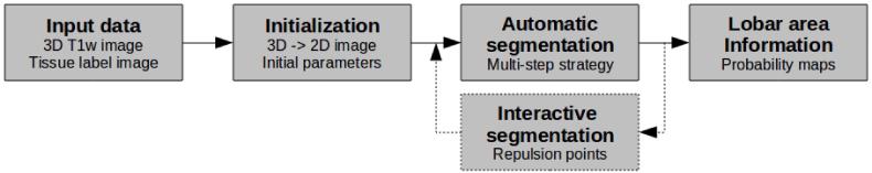
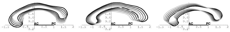
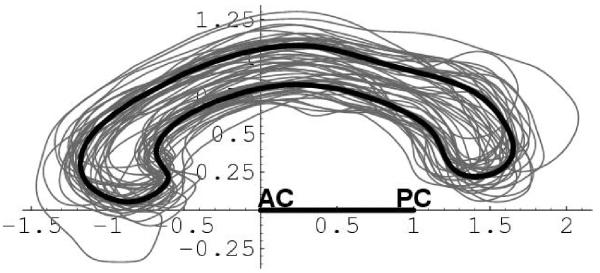
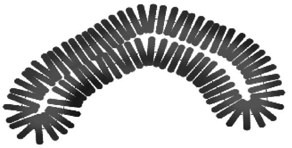
The corpus callosum (CC) is a structure of interest in many neuroimaging studies of neuro-developmental pathology such as autism. It plays an integral role in relaying sensory, motor and cognitive information from homologous regions in both hemispheres. We have developed a framework that allows automatic segmentation of the corpus callosum and its lobar subdivisions. Our approach employs constrained elastic deformation of flexible Fourier contour model, and is an extension of Szekely's 2D Fourier descriptor based Active Shape Model. The shape and appearance model, derived from a large mixed population of 150+ subjects, is described with complex Fourier descriptors in a principal component shape space. Using MNI space aligned T1w MRI data, the CC segmentation is initialized on the mid-sagittal plane using the tissue segmentation. A multi-step optimization strategy, with two constrained steps and a final unconstrained step, is then applied. If needed, interactive segmentation can be performed via contour repulsion points. Lobar connectivity based parcellation of the corpus callosum can finally be computed via the use of a probabilistic CC subdivision model. Our analysis framework has been integrated in an open-source, end-to-end application called CCSeg both with a command line and Qt-based graphical user interface (available on NITRC). A study has been performed to quantify the reliability of the semi-automatic segmentation on a small pediatric dataset. Using 5 subjects randomly segmented 3 times by two experts, the intra-class correlation coefficient showed a superb reliability (0.99). CCSeg is currently applied to a large longitudinal pediatric study of brain development in autism.
Keywords: Fourier coefficient; corpus callosum; segmentation; shape model.
Figures

References
-
- Thompson P, Giedd J, Woods R, MacDonald D, Evans A, Toga A. Growth patterns in the developing brain detected by using continuum mechanical tensor maps. Nature. 2000;6774:190–3. - PubMed
-
- Kass M, Witkin A, Terzopoulos D. Snakes: Active contour models. International Journal of Computer Vision. 1988;1(4):321–331.
-
- Cootes TF, Hill A, Taylor CJ, Haslam J. The use of active shape models for locating structures in medical images. IPMI’93. 1993:33–47.
-
- Szekely G, Kelemen A, Brechbuhler C, Gerig G. Segmentation of 2-d and 3-d objects from mri volume data using constrained elastic deformations of flexible fourier contour and surface models. Medical Image Analysis. 1996;1(1):19–34. - PubMed
Grants and funding
LinkOut - more resources
Full Text Sources