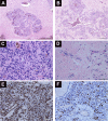
Rapid growth of an orbital hemangiopericytoma with atypical histopathological findings
- PMID: 24353402
- PMCID: PMC3862697
- DOI: 10.2147/OPTH.S47901
Rapid growth of an orbital hemangiopericytoma with atypical histopathological findings
Abstract
Hemangiopericytoma is a rare vascular tumor that originates from pericytes. The orbit is a rare location for this particular tumor, and corresponds to 0.8% to 3% of all primary orbital tumors. We report a case of a hemangiopericytoma in a 45-year-old man that had an unusual presentation, as a rapidly growing mass in the anterior right inferior orbit. Given that there are no clinical or radiological signs pathognomonic of this tumor, a careful histopathological examination is necessary to confirm the diagnosis. In our case, it presented also with unusual histopathological findings. The clinical features, radiological findings, differential diagnosis and treatment of this challenging entity are reviewed in this case report.
Keywords: hemangiopericytoma; orbit; tumor.
Figures


Similar articles
-
Orbital solitary fibrous tumors: a multi-centered histopathological and immunohistochemical analysis with radiological description.Ann Saudi Med. 2020 May-Jun;40(3):227-233. doi: 10.5144/0256-4947.2020.227. Epub 2020 Jun 4. Ann Saudi Med. 2020. PMID: 32493043 Free PMC article.
-
Orbital solitary fibrous tumor: encompassing terminology for hemangiopericytoma, giant cell angiofibroma, and fibrous histiocytoma of the orbit: reappraisal of 41 cases.Hum Pathol. 2011 Jan;42(1):120-8. doi: 10.1016/j.humpath.2010.05.021. Epub 2010 Nov 5. Hum Pathol. 2011. PMID: 21056898
-
Very rare localization of a retroperitoneal hemangiopericytoma revealed by lumbosciatalgia: A case report.Int J Surg Case Rep. 2018;53:127-131. doi: 10.1016/j.ijscr.2018.10.056. Epub 2018 Oct 30. Int J Surg Case Rep. 2018. PMID: 30391737 Free PMC article.
-
Orbital hemangiopericytoma: CT, MR, and angiographic findings.Comput Med Imaging Graph. 1994 May-Jun;18(3):217-22. doi: 10.1016/0895-6111(94)90033-7. Comput Med Imaging Graph. 1994. PMID: 8025890 Review.
-
Meningeal solitary fibrous tumor as an unusual cause of expohthalmos: case report and review of the literature.Neurosurgery. 2001 Jun;48(6):1362-6. doi: 10.1097/00006123-200106000-00039. Neurosurgery. 2001. PMID: 11383743 Review.
Cited by
-
Rare Diseases of the Orbit.Laryngorhinootologie. 2021 Apr;100(S 01):S1-S79. doi: 10.1055/a-1384-4641. Epub 2021 Apr 30. Laryngorhinootologie. 2021. PMID: 34352903 Free PMC article. Review.
-
Sacro-anterior haemangiopericytoma: a case report.Cancer Biol Med. 2014 Jun;11(2):139-43. doi: 10.7497/j.issn.2095-3941.2014.02.010. Cancer Biol Med. 2014. PMID: 25009757 Free PMC article.
-
A Clinical Case of 5 Times Irradiated Recurrent Orbital Hemangiopericytoma.Case Rep Oncol. 2021 Feb 25;14(1):78-84. doi: 10.1159/000513030. eCollection 2021 Jan-Apr. Case Rep Oncol. 2021. PMID: 33776686 Free PMC article.
-
Giant Perivascular Epithelioid Cell Tumor of the Orbit: A Clinicopathological Analysis and Review of the Literature.Ocul Oncol Pathol. 2018 Sep;4(5):272-279. doi: 10.1159/000484425. Epub 2018 Feb 6. Ocul Oncol Pathol. 2018. PMID: 30320097 Free PMC article.
References
-
- Zimmermann KW. Der feinere Bau der Blutcapillaren. [The minute structure of blood capillaries.] Zeitschrift für Anatomie und Entwick-lungsgeschichte. 1923;68(1):29–109.
-
- Ide F, Obara K, Mishima K, Saito I, Kusama K. Ultrastructural spectrum of solitary fibrous tumor: a unique perivascular tumor with alternative lines of differentiation. Virchows Arch. 2005;446:646–652. - PubMed
-
- Henderson JW, Farrow GM. Primary orbital hemangiopericytoma. An aggressive and potentially malignant neoplasm. Arch Ophthalmol. 1978;96:666–673. - PubMed
-
- Croxatto JO, Font RL. Hemangiopericytoma of the orbit: a clinicopathologic study of 30 cases. Hum Pathol. 1982;13:210–218. - PubMed
-
- Tsai CC, Kau HC, Chen SJ, Hsu WM, Wu JS. Primary orbital hemangiopericytoma: a case report. Zhonghua Yi Xue Za Zhi (Taipei) 1997;59:382–385. - PubMed
Publication types
LinkOut - more resources
Full Text Sources
Other Literature Sources

