

APLP2 regulates the expression of MHC class I molecules on irradiated Ewing's sarcoma cells
- PMID: 24353913
- PMCID: PMC3862638
- DOI: 10.4161/onci.26293
APLP2 regulates the expression of MHC class I molecules on irradiated Ewing's sarcoma cells
Abstract
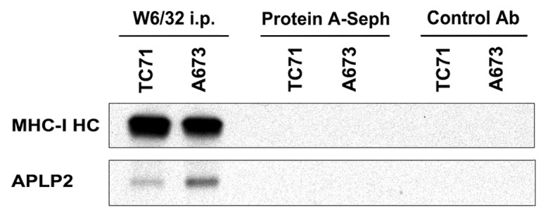
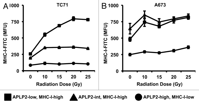
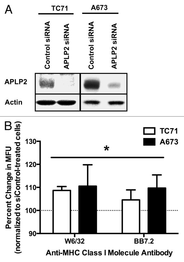
Ewing's sarcoma (EWS) is a pediatric cancer that is conventionally treated by surgery, chemotherapy, and radiation therapy. Innovative immunotherapies to treat EWS are currently under development. Unfortunately for EWS patients, when the disease is found to be resistant to current therapeutic approaches, the prognosis is predictably grim. Radiation therapy and immunotherapy could potentially synergize in the eradication of EWS, as some studies have previously shown that irradiation increases the presence of immune receptors, including MHC class I molecules, on the surface of tumor cells. However, EWS cells have been reported to express low levels of MHC class I molecules, a phenotype that would inhibit T-cell mediated lysis. We have previously demonstrated that the transgene-driven overexpression of amyloid β (A4) precursor-like protein 2 (APLP2) reduces the expression of MHC class I molecules on the surface of human cervical carcinoma HeLa cells. We thus examined whether endogenously expressed APLP2 downregulates MHC class I expression on EWS cells, particularly upon irradiation. We found that irradiation induces the relocalization of APLP2 and MHC class I molecules on the surface of EWS cells, redistributing cells from subpopulations with relatively low APLP2 and high MHC class I into subpopulations with relatively high APLP2 and low MHC class I surface expression. Consistent with these findings, the transfection of an APLP2-targeting siRNA into EWS cells increased MHC class I expression on the cell surface. Furthermore, APLP2 was found by co-immunoprecipitation to bind to MHC class I molecules. Taken together, these findings suggest that APLP2 inhibits MHC class I expression on the surface of irradiated EWS cells by a mechanism that involves APLP2/MHC class I interactions. Thus, therapeutic strategies that limit APLP2 expression may boost the ability of T cells to recognize and eradicate EWS in patients.
Keywords: Ewing’s sarcoma; HLA; MHC class I molecule; amyloid β (A4) precursor-like protein 2; immune evasion; immunotherapy; pediatric cancer; radiation therapy.
Figures

Similar articles
-
Specificity of amyloid precursor-like protein 2 interactions with MHC class I molecules.Immunogenetics. 2008 Jun;60(6):303-13. doi: 10.1007/s00251-008-0296-0. Epub 2008 May 2. Immunogenetics. 2008. PMID: 18452037 Free PMC article.
-
Monocyte Maturation Mediators Upregulate CD83, ICAM-1 and MHC Class 1 Expression on Ewing's Sarcoma, Enhancing T Cell Cytotoxicity.Cells. 2021 Nov 8;10(11):3070. doi: 10.3390/cells10113070. Cells. 2021. PMID: 34831294 Free PMC article.
-
Amyloid precursor-like protein 2 association with HLA class I molecules.Cancer Immunol Immunother. 2009 Sep;58(9):1419-31. doi: 10.1007/s00262-009-0657-z. Epub 2009 Jan 31. Cancer Immunol Immunother. 2009. PMID: 19184004 Free PMC article.
-
Current Status of Proteomics in Ewing's Sarcoma.Proteomics Clin Appl. 2019 May;13(3):e1700130. doi: 10.1002/prca.201700130. Epub 2018 Aug 23. Proteomics Clin Appl. 2019. PMID: 29992772 Review.
-
Association of intracellular proteins with folded major histocompatibility complex class I molecules.Immunol Res. 2004;30(2):171-9. doi: 10.1385/IR:30:2:171. Immunol Res. 2004. PMID: 15477658 Review.
Cited by
-
Gamma Irradiation Triggers Immune Escape in Glioma-Propagating Cells.Cancers (Basel). 2022 May 31;14(11):2728. doi: 10.3390/cancers14112728. Cancers (Basel). 2022. PMID: 35681710 Free PMC article.
-
Beta 2-microglobulin regulates amyloid precursor-like protein 2 expression and the migration of pancreatic cancer cells.Cancer Biol Ther. 2019;20(6):931-940. doi: 10.1080/15384047.2019.1580414. Epub 2019 Feb 27. Cancer Biol Ther. 2019. PMID: 30810435 Free PMC article.
-
Novel regulators of PrPC biosynthesis revealed by genome-wide RNA interference.PLoS Pathog. 2021 Oct 27;17(10):e1010013. doi: 10.1371/journal.ppat.1010013. eCollection 2021 Oct. PLoS Pathog. 2021. PMID: 34705895 Free PMC article.
-
Sex-dependent effects of amyloid precursor-like protein 2 in the SOD1-G37R transgenic mouse model of MND.Cell Mol Life Sci. 2021 Oct;78(19-20):6605-6630. doi: 10.1007/s00018-021-03924-5. Epub 2021 Sep 2. Cell Mol Life Sci. 2021. PMID: 34476545 Free PMC article.
-
Advances in Hypofractionated Irradiation-Induced Immunosuppression of Tumor Microenvironment.Front Immunol. 2021 Jan 25;11:612072. doi: 10.3389/fimmu.2020.612072. eCollection 2020. Front Immunol. 2021. PMID: 33569059 Free PMC article. Review.
References
Publication types
Grants and funding
LinkOut - more resources
Full Text Sources
Other Literature Sources
Research Materials