Primary cerebellar tuberculoma in Arnold-Chiari malformation mimicking posterior cranial fossa tumor: the first report
- PMID: 24353933
- PMCID: PMC3864413
- DOI: 10.1055/s-0031-1296052
Primary cerebellar tuberculoma in Arnold-Chiari malformation mimicking posterior cranial fossa tumor: the first report
Abstract
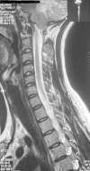
Chiari malformations are a congenital heterogeneous group of disorders characterized by anatomic anomalies of the cerebellum, brain stem, and craniocervical junction associated with downward displacement of the cerebellum, alone or with lower medulla, into the cervical spine canal. The patient was a 23-year-old woman, a known case of Arnold-Chiari malformation with peripheral neuropathy and muscular atrophy, who presented with headache, drowsiness, decreased vision, and severe gait dysfunction lasting for several years. Brain magnetic resonance imaging confirmed a hypointense signal mass in the left hemisphere of the cerebellum causing mass effects on the fourth ventricle, which shifted it, accompanied with dilation of third and lateral ventricles.
Keywords: Arnold-Chiari malformation; cerebellar tuberculoma; cranial fossa tumor.
Figures


Similar articles
-
Posterior cranial fossa box expansion leads to resolution of symptomatic cerebellar ptosis following Chiari I malformation repair.J Craniofac Surg. 2007 Mar;18(2):274-80. doi: 10.1097/scs.0b013e31802c05ab. J Craniofac Surg. 2007. PMID: 17414275
-
Chiari Malformation Type 2.2024 Feb 25. In: StatPearls [Internet]. Treasure Island (FL): StatPearls Publishing; 2025 Jan–. 2024 Feb 25. In: StatPearls [Internet]. Treasure Island (FL): StatPearls Publishing; 2025 Jan–. PMID: 32491430 Free Books & Documents.
-
Analysis of the volumes of the posterior cranial fossa, cerebellum, and herniated tonsils using the stereological methods in patients with Chiari type I malformation.ScientificWorldJournal. 2012;2012:616934. doi: 10.1100/2012/616934. Epub 2012 May 2. ScientificWorldJournal. 2012. PMID: 22629166 Free PMC article.
-
Neuroradiological diagnosis of Chiari malformations.Neurol Sci. 2011 Dec;32 Suppl 3:S283-6. doi: 10.1007/s10072-011-0695-0. Neurol Sci. 2011. PMID: 21800079 Review.
-
Neuroepithelial cyst of the posterior fossa: two case reports with radiologic-pathologic correlation.Can Assoc Radiol J. 1996 Apr;47(2):126-31. Can Assoc Radiol J. 1996. PMID: 8612085 Review.
Cited by
-
Tubercular cerebellitis, identified through an expansive process: A case report.Radiol Case Rep. 2024 Aug 10;19(11):4788-4793. doi: 10.1016/j.radcr.2024.07.110. eCollection 2024 Nov. Radiol Case Rep. 2024. PMID: 39228930 Free PMC article.
-
Giant Tuberculomas of Brain: Rare Neoplastic Mimic.J Pediatr Neurosci. 2020 Jul-Sep;15(3):204-213. doi: 10.4103/jpn.JPN_78_19. Epub 2020 Nov 6. J Pediatr Neurosci. 2020. PMID: 33531933 Free PMC article.
References
-
- Sarnat HB. Disorders of segmentation of the neural tube: Chiari malformations. Handb Clin Neurol. 2008;87:89–103. - PubMed
-
- Carmel PW, Markesbery WR. Early descriptions of the Arnold-Chiari malformation. The contribution of John Cleland. J Neurosurg. 1972;37:543–547. - PubMed
-
- Schijman E. History, anatomic forms, and pathogenesis of Chiari I malformations. Childs Nerv Syst. 2004;20:323–328. - PubMed
-
- Speer MC, Enterline DS, Mehltretter L. et al.Chiari type I malformation with or without syringomyelia: prevalence and genetics. J Genet Couns. 2003;12:297–311. - PubMed
LinkOut - more resources
Full Text Sources

