

Bilateral three-level lumbar spondylolysis repaired by hook-screw technique
- PMID: 24353947
- PMCID: PMC3864455
- DOI: 10.1055/s-0032-1307255
Bilateral three-level lumbar spondylolysis repaired by hook-screw technique
Abstract
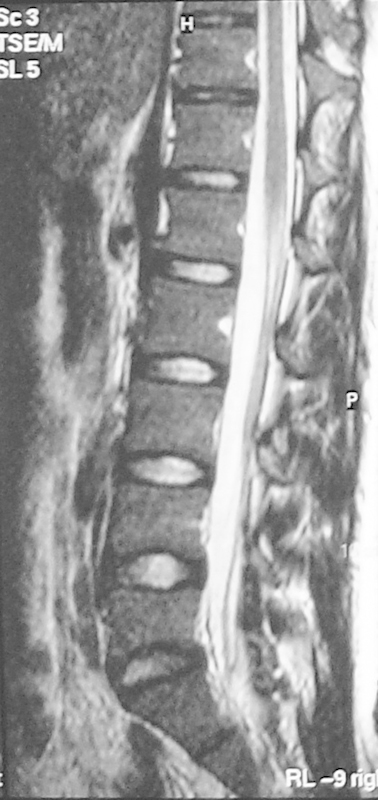
We report a case of bilateral three-level lumbar spondylolysis that was directly repaired by use of hook-screw technique. The patient complained of low back pain for 2 years that progressively worsened and was exacerbated with standing and walking. He also mentioned bilateral sciatalgia. The neurological examination was normal. Interestingly, we found bilateral lumbar spondylolysis in L3, L4, and L5 levels in imaging studies. After proving that spondylolysis was the source of the low back pain by local anesthetic agent injection, we used a direct technique for correction of spondylolysis by use of a hook-screw device plus decortications of lysis area and iliac crest autograft. We assessed the patient after surgery to evaluate pain recovery and fusion rate. The results were favorable and proved the efficacy of the hook-screw technique for treatment of symptomatic multilevel lumbar spondylolysis.
Keywords: hook-screw; low back pain; spondylolysis.
Figures


Similar articles
-
Direct repair of spondylolysis by TSRH's hook plus screw fixation and bone grafting: biomechanical study and clinical report.Arch Orthop Trauma Surg. 2010 Feb;130(2):209-15. doi: 10.1007/s00402-009-0897-6. Epub 2009 May 14. Arch Orthop Trauma Surg. 2010. PMID: 19440723
-
[Direct repair of adolescent lumbar spondylolysis using a pedicle screw-laminar hook system by paramedian approach].Zhongguo Gu Shang. 2011 Aug;24(8):687-9. Zhongguo Gu Shang. 2011. PMID: 21928681 Chinese.
-
Surgical treatment of four segment lumbar spondylolysis: A case report.World J Clin Cases. 2021 Jun 16;9(17):4408-4414. doi: 10.12998/wjcc.v9.i17.4408. World J Clin Cases. 2021. PMID: 34141808 Free PMC article.
-
Double-level lumbar spondylolysis and spondylolisthesis: A retrospective study.J Orthop Surg Res. 2018 Mar 16;13(1):55. doi: 10.1186/s13018-018-0723-3. J Orthop Surg Res. 2018. PMID: 29548343 Free PMC article. Review.
-
[Clinical research progress of direct surgical repair of lumbar spondylolysis in young patients].Zhongguo Xiu Fu Chong Jian Wai Ke Za Zhi. 2013 Jan;27(1):106-9. Zhongguo Xiu Fu Chong Jian Wai Ke Za Zhi. 2013. PMID: 23427504 Review. Chinese.
Cited by
-
Surgical Management of 3-Level Lumbar Spondylolyses.Medicine (Baltimore). 2015 Jul;94(27):e1127. doi: 10.1097/MD.0000000000001127. Medicine (Baltimore). 2015. PMID: 26166116 Free PMC article.
-
Congenital cervical isthmic spondylolisthesis: A case report.Surg Neurol Int. 2019 Apr 24;10:57. doi: 10.25259/SNI-92-2019. eCollection 2019. Surg Neurol Int. 2019. PMID: 31528395 Free PMC article.
References
-
- Buck J E. Direct repair of the defect in spondylolisthesis. Preliminary report. J Bone Joint Surg Br. 1970;52:432–437. - PubMed
-
- Chang J H, Lee C H, Wu S S, Lin L C. Management of multiple level spondylolysis of the lumbar spine in young males: a report of six cases. J Formos Med Assoc. 2001;100:497–502. - PubMed
-
- Eingorn D, Pizzutillo P D. Pars interarticularis fusion of multiple levels of lumbar spondylolysis. A case report. Spine. 1985;10:250–252. - PubMed
-
- Nozawa S, Shimizu K, Miyamoto K, Tanaka M. Repair of pars interarticularis defect by segmental wire fixation in young athletes with spondylolysis. Am J Sports Med. 2003;31:359–364. - PubMed
LinkOut - more resources
Full Text Sources
