

Microendoscopic Decompression Surgery for Lumbar Spinal Canal Stenosis via the Paramedian Approach: Preliminary Results
- PMID: 24353952
- PMCID: PMC3864472
- DOI: 10.1055/s-0032-1319774
Microendoscopic Decompression Surgery for Lumbar Spinal Canal Stenosis via the Paramedian Approach: Preliminary Results
Abstract
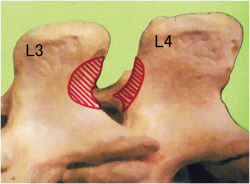
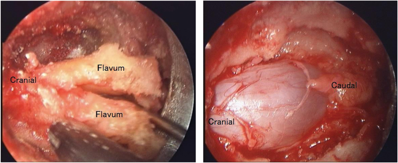
The objective of this study was to evaluate the efficacy of a microendoscopic spinal decompression surgical technique using a novel approach for the treatment of lumbar spinal canal stenosis (LSCS). The following modifications were made to the conventional microendoscopic bilateral decompression via the unilateral approach: the base of the spinous process was first resected partially to secure a working space, so as not to separate the spinous process from the lamina. The tip of the tubular retractor was placed at the midline of the lamina, where laminectomy was performed microendoscopically. A total of 126 stenotic levels were decompressed in 70 patients. The mean operating time per level was 77.0 minutes, and the mean intraoperative blood loss per level was 15.0 mL. There were no dural tears or neurological injuries intraoperatively. Fracture of the spinous process was detected postoperatively in two patients, both of whom were asymptomatic. All patients could be followed up for at least 12 months. Their median Japanese Orthopaedic Association (JOA) score improved significantly from 16 points preoperatively to 27.5 points after the surgery (p < 0.001). The case series showed that the modifications of the technique improved the safety and ease of performance of the microendoscopic decompression surgery for LSCS.
Keywords: kissing spine; lumbar spinal canal stenosis; microendoscopic surgery; minimally invasive surgery; posterior decompression; spinous process.
Figures

References
-
- Foley K T, Smith M M. Microendoscopic discectomy. Tech Neurosurg. 1997;3:301–307.
-
- Yoshida M. Kyoto: Kimpodo; 2005. Microendoscopic Discectomy; pp. 63–83.
-
- Watanabe K, Hosoya T, Shiraishi T, Matsumoto M, Chiba K, Toyama Y. Lumbar spinous process-splitting laminectomy for lumbar canal stenosis. Technical note. J Neurosurg Spine. 2005;3(5):405–408. - PubMed
-
- Hatta Y, Shiraishi T, Sakamoto A. et al.Muscle-preserving interlaminar decompression for the lumbar spine: a minimally invasive new procedure for lumbar spinal canal stenosis. Spine. 2009;34(8):E276–E280. - PubMed
-
- Yanagisawa K, Mikami Y, Hatta Y, Hase H, Koyama K, Kubo T. New microendoscopic decompression method via midline interlaminar approach for lumbar canal stenosis: microendoscopic muscle preserving interlaminar decompression; ME-MILD. Surgical Technique for Spine and Spinal Nerves. 2006;8:105–108.
LinkOut - more resources
Full Text Sources
Other Literature Sources
