Acute Schmorl's Node during Strenuous Monofin Swimming: A Case Report and Review of the Literature
- PMID: 24353963
- PMCID: PMC3864465
- DOI: 10.1055/s-0032-1307262
Acute Schmorl's Node during Strenuous Monofin Swimming: A Case Report and Review of the Literature
Abstract
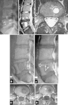
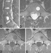
Study Design This case report describes an acute Schmorl's node (SN) in an elite monofin athlete during exercise. The patient presented with severe back pain and leg numbness and was managed successfully with conservative treatment. Objective The aim of our communication was to describe a rare presentation of a common pathological condition during an intense sport. Background Swimming is not generally considered to be a sport activity that leads to spinal injuries. SNs are usually asymptomatic lesions, incidentally found on imaging studies. There is no correlation between swimming and symptomatic SN formation. Case Report A 16-year-old monofin elite athlete suffered from an acute nonradiating back pain during extreme exercise. His back pain was associated with a fracture of the superior L5 end plate and an acute SN at the L5 vertebral body with perilesional bone marrow edema. The pain resolved with nonsteroidal anti-inflammatory drugs and bed rest. The athlete had an excellent outcome and returned to his training activities 6 months after his incident. Conclusion SN should be considered in the differential diagnosis of severe back pain, especially in sport-related injuries. SNs present with characteristic imaging findings. Due to the benign nature of these lesions, surveillance-only management may be the best course of action.
Keywords: MR imaging/diagnosis; acute Schmorl's node/giant; conservative treatment; monofin swimming.
Conflict of interest statement
Figures


Similar articles
-
Acute Lumbar Schmorl's Node Following Chiropractic Adjustment.Cureus. 2022 May 22;14(5):e25209. doi: 10.7759/cureus.25209. eCollection 2022 May. Cureus. 2022. PMID: 35746996 Free PMC article.
-
Schmorl's Node: An Uncommon Case of Back Pain and Radiculopathy.Orthop Rev (Pavia). 2022 Apr 25;14(3):33641. doi: 10.52965/001c.33641. eCollection 2022. Orthop Rev (Pavia). 2022. PMID: 35775032 Free PMC article.
-
Relationship of Schmorl's nodes to vertebral body endplate fractures and acute endplate disk extrusions.AJNR Am J Neuroradiol. 2000 Feb;21(2):276-81. AJNR Am J Neuroradiol. 2000. PMID: 10696008 Free PMC article.
-
Schmorl's nodes.Eur Spine J. 2012 Nov;21(11):2115-21. doi: 10.1007/s00586-012-2325-9. Epub 2012 Apr 28. Eur Spine J. 2012. PMID: 22544358 Free PMC article. Review.
-
Percutaneous Vertebroplasty for Symptomatic Schmorl's Nodes: 11 Cases with Long-term Follow-up and a Literature Review.Pain Physician. 2017 Feb;20(2):69-76. Pain Physician. 2017. PMID: 28158154 Review.
Cited by
-
Infected Schmorl's node: a case report.BMC Musculoskelet Disord. 2020 May 2;21(1):280. doi: 10.1186/s12891-020-03276-4. BMC Musculoskelet Disord. 2020. PMID: 32359347 Free PMC article.
-
Acute Lumbar Schmorl's Node Following Chiropractic Adjustment.Cureus. 2022 May 22;14(5):e25209. doi: 10.7759/cureus.25209. eCollection 2022 May. Cureus. 2022. PMID: 35746996 Free PMC article.
-
A Retrospective Study of Percutaneous Balloon Kyphoplasty for the Treatment of Symptomatic Schmorl's Nodes: 5-Year Results.Med Sci Monit. 2017 Jun 13;23:2879-2889. doi: 10.12659/msm.904802. Med Sci Monit. 2017. PMID: 28607331 Free PMC article.
References
-
- Mok FP, Samartzis D, Karppinen J, Luk KD, Fong DY, Cheung KM. ISSLS prize winner: prevalence, determinants, and association of Schmorl nodes of the lumbar spine with disc degeneration: a population-based study of 2449 individuals. Spine. 2010;35:1944–1952. - PubMed
-
- Dar G, Peleg S, Masharawi Y, Steinberg N, May H, Hershkovitz I. Demographical aspects of Schmorl nodes: a skeletal study. Spine. 2009;34:E312–E315. - PubMed
-
- Boden BP Jarvis CG Spinal injuries in sports Neurol Clin 20082663–78., viii - PubMed
-
- Karantanas A. Berlin, Heidelberg: Springer-Verlag; 2011. Common injuries in water sports; pp. 289–316.
-
- Schmorl G. Die pathologische Anatomie der Wirbelsaule. Verh Dtsch Orthop Ges. 1927;21:3–41.
Publication types
LinkOut - more resources
Full Text Sources

