. 2013 Nov 29:1318:251-6.
doi: 10.1016/j.chroma.2013.10.020.

A microchip electrophoresis-mass spectrometric platform for fast separation and identification of enantiomers employing the partial filling technique

A microchip electrophoresis-mass spectrometric platform for fast separation and identification of enantiomers employing the partial filling technique

Xiangtang Li et al. J Chromatogr A. .

Abstract

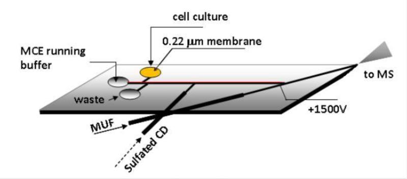
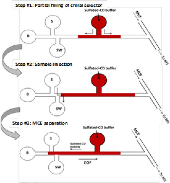
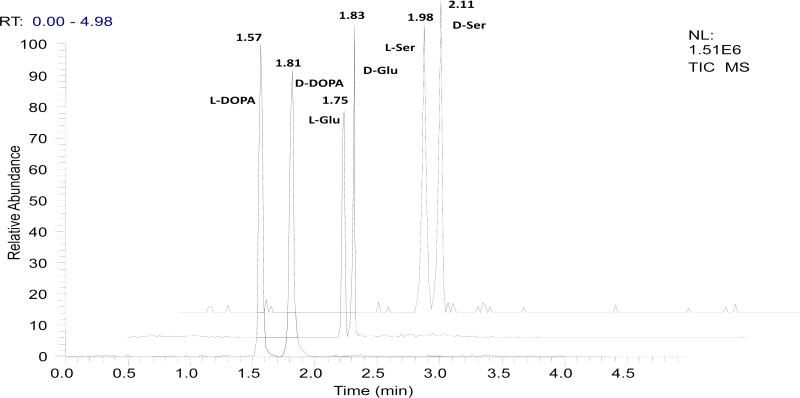
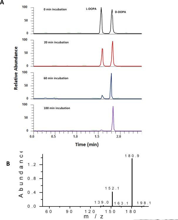
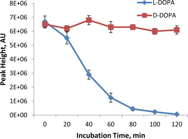
A microchip electrophoresis-mass spectrometric (MCE-MS) method was developed for fast chiral analysis. The proposed MCE-MS platform deployed a glass/PDMS hybrid microchip with an easy-to-fabricate monolithic nanoelectrospray emitter. Enantiomeric MCE separation was achieved by means of the partial filling technique. A novel chip design with an arm channel connecting to the middle of the MCE separation channel for delivering the chiral selector was tested and proven valid. Enantiomeric separation of3.4-dihydroxyphenylalanine (DOPA), glutamic acid (Glu), and serine (Ser), the selected test compounds,were achieved within 130 s with resolution values (R(s)) of 2.4, 1.1, and 1.0, respectively. The proposed chiral MCE-MS assay was sensitive and had detection limits of 43 nM for l-DOPA and 47 nM for d-DOPA.The analytical platform was well suited for studies of stereochemical preference in living cells because it integrated cell culture, sample injection, chiral separation, and MS detection into a single platform.Metabolism of DOPA in human SH-SY5Y neuronal cells was studied as a model system. On-chip incubation of SH-SY5Y cells with racemic DOPA was carried out, and the incubation solution was injected and in-line assayed at time intervals. It was found that l-DOPA concentration decreased gradually as incubation time increased while the concentration of coexisting d-DOPA remained constant. The results firmly indicated that SH-SY5Y cells metabolized l-DOPA effectively while left d-DOPA intact.


Figures

Fig 1
Fig 1
Microchip design used in the proposed MCE-MS platform (channels were 60 μm wide × 20 μm deep).
Fig 2
Fig 2
A schematic illustration of the proposed partial filling chiral MCE-MS analysis with sulfated β-CD as chiral selector. The approach can be easily executed in a microfluidic chip. Delivery of chiral selector is done preferably by using a gas-tight syringe (hydrodynamically) over by applying an electrical potential (electrokinetically).
Fig 3
Fig 3
TIC electropherograms from the proposed chiral MCE-MS separation of neuroactive compounds, i.e. DOPA, glutamic acid (Glu), and serine (Ser). [test compound] = 100 μM (each enantiomer). Chiral MCE conditions: MCE separation channel, 4 cm long × 60 μm wide × 20 μm deep; electrokinetic sample injection, 15 s at 600V; 35nL chiral selector solution infused by a syringe pump; separation voltage, 3850V; electrospray voltage,1500V; MCE running buffer, 15mM ammonium acetate/ acetic acid buffer (pH 5.5) /methanol (1:1); chiral selector solution, the MCE running buffer containing 15 mM sulfated β-CD; MUF, the MCE running buffer at a flow rate of 100 nL /min.
Fig 4
Fig 4
Electropherograms obtained from studying DOPA metabolism in SH-SY5Y neuronal cells: (A) TICs of m/z 198 from incubation solution injected at different times, and (B) MS/MS spectrum of the peaks, confirming DOPA identity. MCE-MS conditions were as in Fig. 3. Peak height of L-DOPA decreases gradually as incubation time increases while that of co-existing D-DOPA remains unchanged.
Fig 5
Fig 5
Metabolic trend curves of L-DOPA and D-DOPA (at 50 μM each) in incubation with SH-SY5Y neuronal cells as measured by the proposed chiral MCE-MS method (mean ± SD, n=3).

References

    1. Taniguchi K, Maeda R, Ando T, Okumura T, Nakazawa N, Hatori R, Nakamura M, Hozumi S, Fujiwara H, Matsuno K. Science. 2011;333:339. - PubMed
    1. Wan LQ, Ronaldson K, Park M, Taylor G, Zhang Y, Gimble JM, Vunjak-Novakovic G. Proc Natl Acad Sci U S A. 2011;108:12295. - PMC - PubMed
    1. Zask A, Murphy J, Ellestad GA. Chirality. 2013;25:265. - PubMed
    1. Caglioti L, Micskei K, Palyi G. Chirality. 2011;23:65. - PubMed
    1. Wan LQ, Ronaldson K, Guirguis M, Vunjak-Novakovic G. Stem Cell Res Ther. 2013;4:24. - PMC - PubMed

Publication types

LinkOut - more resources