Review
. 2014 Mar;171(5):1180-94.
doi: 10.1111/bph.12544.

Biased signalling and proteinase-activated receptors (PARs): targeting inflammatory disease

Affiliations
Review

Biased signalling and proteinase-activated receptors (PARs): targeting inflammatory disease

M D Hollenberg et al. Br J Pharmacol. 2014 Mar.

Abstract

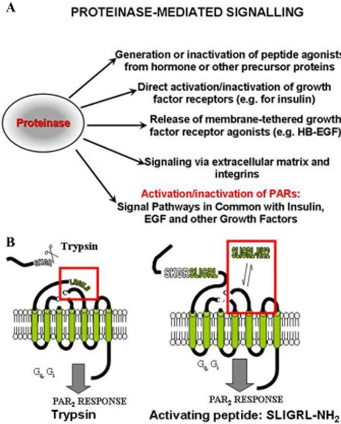
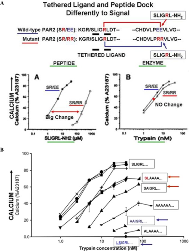
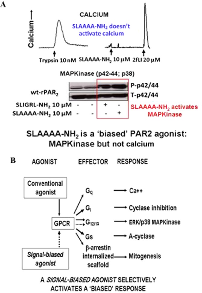
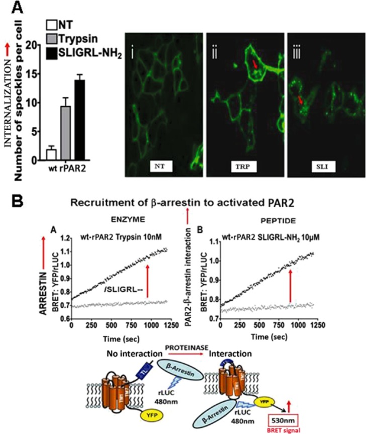
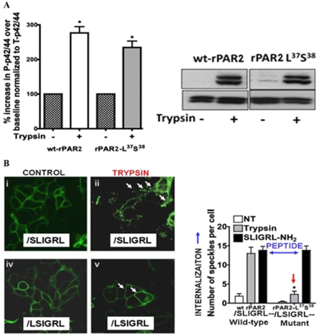
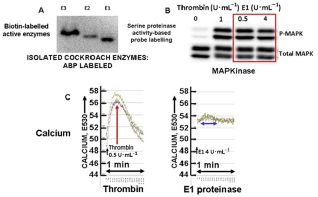
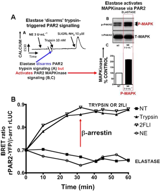
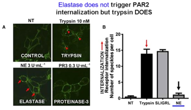
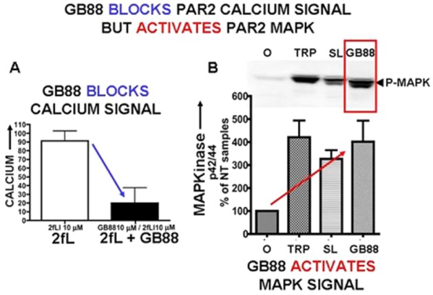
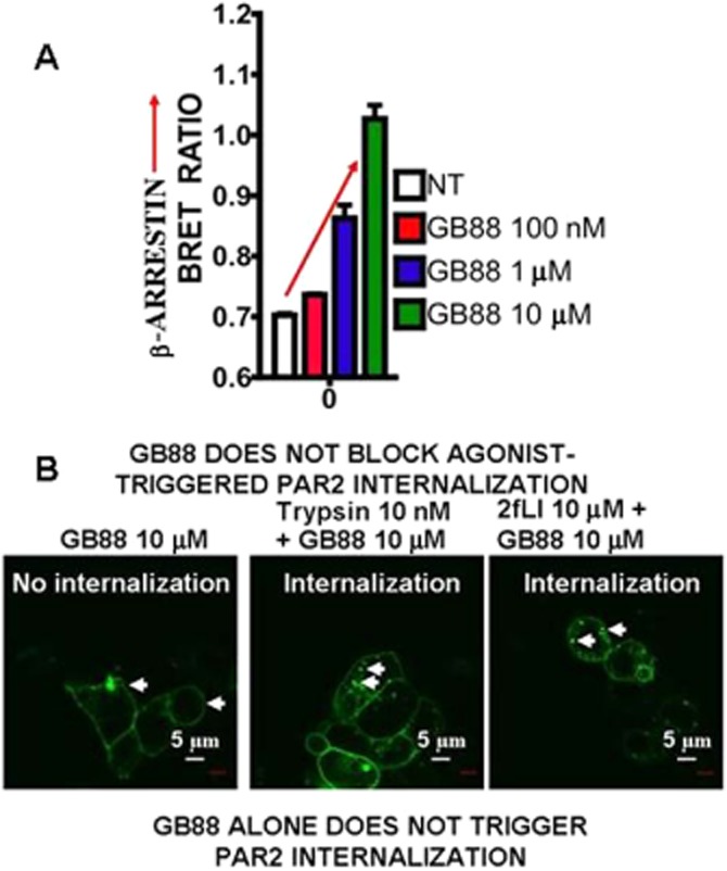
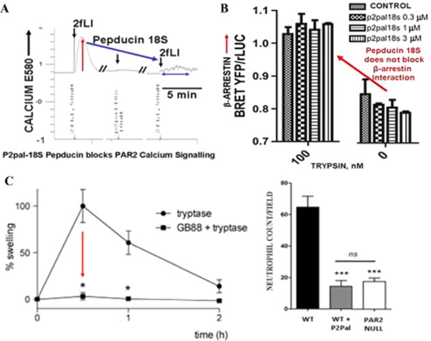
Although it has been known since the 1960s that trypsin and chymotrypsin can mimic hormone action in tissues, it took until the 1990s to discover that serine proteinases can regulate cells by cleaving and activating a unique four-member family of GPCRs known as proteinase-activated receptors (PARs). PAR activation involves the proteolytic exposure of its N-terminal receptor sequence that folds back to function as a 'tethered' receptor-activating ligand (TL). A key N-terminal arginine in each of PARs 1 to 4 has been singled out as a target for cleavage by thrombin (PARs 1, 3 and 4), trypsin (PARs 2 and 4) or other proteases to unmask the TL that activates signalling via Gq , Gi or G12 /13 . Similarly, synthetic receptor-activating peptides, corresponding to the exposed 'TL sequences' (e.g. SFLLRN-, for PAR1 or SLIGRL- for PAR2) can, like proteinase activation, also drive signalling via Gq , Gi and G12 /13 , without requiring receptor cleavage. Recent data show, however, that distinct proteinase-revealed 'non-canonical' PAR tethered-ligand sequences and PAR-activating agonist and antagonist peptide analogues can induce 'biased' PAR signalling, for example, via G12 /13 -MAPKinase instead of Gq -calcium. This overview summarizes implications of this 'biased' signalling by PAR agonists and antagonists for the recognized roles the PARs play in inflammatory settings.

Keywords: GPCR; activated protein-C; biased signalling; protease; proteinase-activated receptor; thrombin.


Figures

Figure 1
Figure 1
Proteinase-mediated signalling (A) and the mechanisms of PAR activation (B). (A, upper) The diagram shows five distinct ways by which proteinases can cause cell signals, ranging from the generation or degradation of active peptides (top) to the activation of PARs (bottom). (B, lower) The scheme shows the proteolytic activation of PARs either by the unmasking of a receptor-tethered activating ligand (left: SLIGRL— for PAR2) or by a synthetic peptide with a sequence in common with the revealed ‘TL’ (right: SLIGRL-NH2).
Figure 2
Figure 2
Differences in signalling by PAR2-activating peptide versus TL in a mutant PAR2 ‘SR/RR’ receptor (A, upper, panels A and B) and minimal TL sequence needed for calcium signalling (B, lower). (A, upper) The scheme at the top shows the wild-type receptor sequence (PAR2: SR/EE), with a TL sequence, ‘SLIGRL---) thought to interact with the ‘PEE’ sequence in extracellular loop-2. This wild-type receptor is compared with a mutant PAR2 having the same ‘TL’ (SLIGR---) but a mutated ‘PRR’ sequence in extracellular loop 2. The upper-left figure A shows calcium signalling stimulated by the PAR2-activating peptide, SLIGRL-NH2 in the wild-type (SR/EE), compared with the mutant (SR/RR) receptor, demonstrating a marked rightward shift in the concentration-effect curve for the mutant (SR/RR) versus the wild-type receptor (SR/EE). In contrast, the upper-right panel B shows there is no difference between the wild-type (SR/EE) versus mutant (SR/RR) receptor for activation by the trypsin-revealed TL. Thus, the receptor-docking sites for signalling by the synthetic PAR-activating peptide and the proteinase-unmasked TLs differ. Adapted from Al-Ani et al., with permission. (B, lower) A minimal PAR2-TL sequence (SL----) is required for calcium signalling. The proteinase-revealed ‘TL’ sequence of PAR2 was mutated so that trypsin cleavage unmasked different mutated ‘TL’ sequences with alanine substitutions (sequences shown in boxes) in the first six amino acids. When revealed, the mutated TL sequences, SLAAAA--- and SAIGRL--- were able to stimulate calcium signalling (red arrows, upper curves), whereas the sequences, AAIGRL--- and LSIGRL--- did not (blue arrows, bottom). Adapted from Al-Ani et al., , with permission.
Figure 3
Figure 3
Biased PAR2 signalling by the PAR-activating peptide, SLAAAA-NH2 (A) and a scheme (B) that illustrates the ‘mobile’ or ‘floating’ receptor model of signal transduction. (A) Trypsin (A: left tracing) and the PAR2-activating peptide (2-furoyl-LIGRLO-NH2: right tracing, far-right) both stimulate PAR2 calcium signalling, but the activating peptide with multiple alanine substitutions (SLAAAA-NH2) fails to do so (right tracing, first agonist). However, SLAAAA-NH2, like SLIGRL-NH2, does nonetheless activate MAPKinase signalling, acting as a ‘biased’ PAR2 agonist (lower Western blot signal, red rectangle, panel A). Adapted from Ramachandran et al., , with permission. (B) This ‘biased’ signalling by PAR2 is illustrated by the scheme (lower panel B). Thus, an individual GPCR capable of coupling to multiple G-proteins, as well as generating a β-arrestin internalized signalling scaffold, can in principle be activated selectively by a ‘biased’ agonist to signal via only one of the available G-proteins (dotted lines).
Figure 4
Figure 4
Monitoring PAR2 internalization and arrestin interaction upon activation. (A) PAR2 internalization (red arrows) after activation by either trypsin (TRP: middle micrograph) or PAR2-activating peptide (SLI: right-hand micrograph). Receptor in the untreated cells (NT) is membrane delimited. Internalization is quantified (left histograms) by morphometric analysis of internalized receptor clusters (green dots, red arrows, micrographs on the right, panel A). (B) Monitoring PAR2-β-arrestin interaction by BRET after activation by the TL revealed by trypsin (left) or by a synthetic PAR-activating peptide (right). PAR2-β-arrestin interactions stimulated by either trypsin (B, panel A) or PAR2-activating peptide (B, panel B) are quantified by measuring the yelllow fluorescent protein (YFP) emission at 540 nm relative to the rLUC signal (YFP/rLUC), caused by BRET (panel B), as shown by the diagram at the bottom of B. Adapted from Ramachandran et al., , with permission.
Figure 5
Figure 5
Biased MAPKinase signalling by a mutated rat PAR2-TL (A) without receptor internalization (B). (A, upper) Activation of PAR2 MAPKinase signalling by the trypsin-exposed mutated TL, //L37S38IGRL--- (rPAR2-L37S38). PAR2 was mutated so that trypsin cleavage (shown by //) at the R36//L37SIGRL--- sequence unmasks the TL sequence, L37S38IGRL--- (rPAR2-L37S38), which activates MAPKinase to the same level as for trypsin-activated wild-type receptor [A, histograms (left) and Western blot (right)]. However, the trypsin-unmasked mutant TL (rPAR2-L37S38) does not activate calcium signalling (Figure 2B, lower panel, blue arrow at the bottom, X-axis). (B) However, activation of the mutant PAR2 (rPAR2-L37S38) by the trypsin-exposed TL, //LSIGRL--- does not trigger receptor internalization (lower-right image, Figure 5B; red arrow, right histograms). In contrast, peptide-driven receptor activation causes internalization of both the wild-type and mutant rPAR2-L37S38 receptors (blue two-way arrow, right-hand histograms, Figure 5B). Adapted from Ramachandran et al., .
Figure 6
Figure 6
Biased PAR1 signalling (MAPKinase, but not calcium) by purified cockroach allergen serine proteinase E1. (A) Biotin-labelling of isolated cockroach proteinases E1, E2 and E3 with the biotin-serine proteinase activity-based probe (ABP). (B) Activation of MAPKinase in PAR1-expressing cells (Western blot signal: P-MAPK) by cockroach proteinase E1 (0.5 and 4 U·mL−1 enzyme activity: red outline) compared with thrombin (1 U·mL−1). (C) Calcium signalling stimulated by thrombin (0.5 U·mL−1: red arrow, left panel E530) but not by E1 proteinase (4 U·mL−1: blue two-way arrow, right panel, E530) at concentrations that nonetheless trigger MAPKinase activation (Figure 6B, red outline and data not shown).
Figure 7
Figure 7
Neutrophil elastase (NE) stimulates ‘biased’ PAR2 signalling (MAPKinase (A, panel B); but not calcium (A, panel A) without triggering a PAR2-β-arrestin interaction (B). (A, panel A) NE, which does not cause a calcium signal on its own, disarms PAR2 preventing trypsin-stimulated (blue arrow) but not peptide-stimulated PAR2 calcium signalling (increase in E530). (A, sections B and C) NE activates PAR2 MAPKinase signalling. (B, bottom panel) Elastase activation of PAR2 does not trigger a β-arrestin interaction (bottom curve:), whereas trypsin and a PAR-activating peptide (2-furoyl-LIGRLO-NH2:) do (red arrow: upper curves). Adapted from Ramachandran R et al., , with permission.
Figure 8
Figure 8
Neutrophil elastase (NE) does not stimulate PAR2 internalization whereas trypsin does. Trypsin-stimulated PAR2 internalization (upper-right micrograph, panel A, red arrows) was quantified by morphometric analysis of internalized receptor clusters (green dots: arrows, quantified in the right filled histogram: panel B, red arrow). No internalization was observed when cells were treated with NE (lower left quadrant image, panel A; right-hand histogram, NE, black arrow, panel B). Adapted from Ramachandran R et al., with permission.
Figure 9
Figure 9
The PAR2 antagonist GB88 (10 μM) blocks calcium signalling in PAR2-expressing KNRK cells (A, left) but activates PAR2 MAPKinase signalling (B, right). (A) Pretreatment of cells with GB88 (10 μM) blocked PAR-agonist (2-furoyl-LIGRLO-NH2, 10 μM)-stimulated calcium signalling (histograms, panel A), but (B) GB88 (3 μM) stimulated MAPKinase signalling (phospho-p42/44; P-MAPK) relative to ‘no-treatment’ (NT) cells [% p42/44 relative to untreated (0) cells: panel B, red arrow] in PAR2-expressing KNRK cells. The level of the GB88-stimulated MAPKinase was equivalent to that triggered by either trypsin (TRP 1 U mL−1; 10 nM) or a PAR2-activating peptide (SL: SLIGRL-NH2: 10 μM). Data from Ramachandran R et al. unpublished experiments, 2013.
None
The PAR2 ligand, GB88 (0.1–10 μM), that stimulates MAPKinase, promotes β-arrestin interactions (A), but does not either cause receptor internalization on its own (B, left panel; GB88 10 μM) or block agonist-triggered PAR2 internalization (B, middle and right panels). (A) The concentration-dependence of GB88-stimulated PAR2 interaction with β-arrestin is shown in panel A (upper). PAR2 internalization caused by either trypsin (10 nM, middle panel, Figure 10B) or a PAR2 peptide agonist (2-furoyl-LIGRLO-NH2: 2fL; 10 μM, right panel, Figure 10B) is shown by the white arrows. The scale marker (white bar) represents 5 microns (μm). Data from Ramachandran R et al. unpublished experiments, 2013.
Figure 11
Figure 11
Pepducin P2pal-18S blocks calcium signalling (A) but does not affect agonist-stimulated PAR2-β-arrestin interactions (B). However, both P2pal-18S and GB88 inhibit inflammation in vivo, despite their ‘biased antagonist action’ (C). (A) Calcium signalling (E580) stimulated by 2-furoyl-LIGRLO-NH2 (2fLI: 50 μM) in PAR2-expressing HEK cells (upward deflection of tracing) is blocked by pretreatment of the cells with P2pal-18S-pepducin (10 μM, arrow on right) (Data from Ramachandran et al. unpublished experiments, 2013). (B) However, treatment of cells with concentrations of pepducin P2pal-18S that block calcium signalling (A, upper) does not affect the trypsin-stimulated interaction of PAR2 with β-arrestin monitored by BRET (panel B, red arrow: histograms on right (Data from Ramachandran et al. unpublished experiments, 2013). (C) Nonetheless, P2pal-18S (C, right panel histogram, WT + P2PAL) blocks paw inflammation neutrophil influx in vivo to the level found in PAR2 knockout mice; and GB88 (10 mg·kg−1 p.o.) also blocks paw oedema in a tryptase model (20 μg per paw) of peripheral paw inflammation (11C, left panel). Recalculated from Sevigny et al., and adapted from Lohman et al. , with permission.

Similar articles

Cited by

References

    1. Adams MN, Ramachandran R, Yau MK, Suen JY, Fairlie DP, Hollenberg MD, et al. Structure, function and pathophysiology of protease activated receptors. Pharmacol Ther. 2011;130:248–282. - PubMed
    1. Al-Ani B, Saifeddine M, Hollenberg MD. Detection of functional receptors for the proteinase-activated-receptor-2-activating polypeptide, SLIGRL-NH2, in rat vascular and gastric smooth muscle. Can J Physiol Pharmacol. 1995;73:1203–1207. - PubMed
    1. Al-Ani B, Wijesuriya SJ, Hollenberg MD. Proteinase-activated receptor 2: differential activation of the receptor by tethered ligand and soluble peptide analogs. J Pharmacol Exp Ther. 2002;302:1046–1054. - PubMed
    1. Al-Ani B, Hansen KK, Hollenberg MD. Proteinase-activated receptor-2: key role of amino-terminal dipeptide residues of the tethered ligand for receptor activation. Mol Pharmacol. 2004;65:149–156. - PubMed
    1. Alexander SPH, Benson HE, Faccenda E, Pawson AJ, Sharman JL, Spedding M, et al. The Concise Guide to PHARMACOLOGY 2013/14: G Protein-Coupled Receptors. Br J Pharmacol. 2013;170:1459–1581. - PMC - PubMed

Publication types

MeSH terms

Substances