. 2014 Feb;91(4):790-804.
doi: 10.1111/mmi.12498. Epub 2014 Jan 7.

The 4-cysteine zinc-finger motif of the RNA polymerase regulator DksA serves as a thiol switch for sensing oxidative and nitrosative stress

Affiliations

The 4-cysteine zinc-finger motif of the RNA polymerase regulator DksA serves as a thiol switch for sensing oxidative and nitrosative stress

Calvin A Henard et al. Mol Microbiol. 2014 Feb.

Abstract

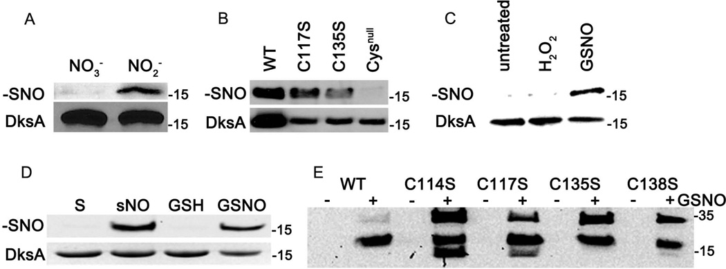
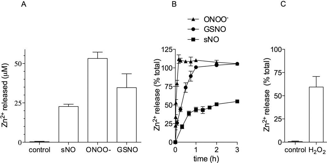
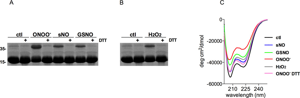
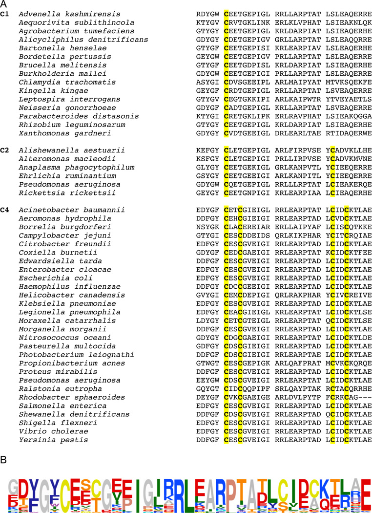
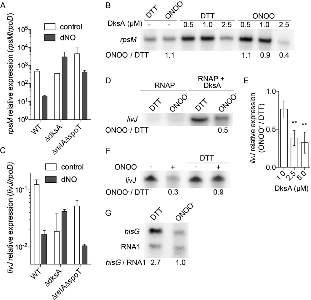
We show that thiols in the 4-cysteine zinc-finger motif of DksA, an RNA polymerase accessory protein known to regulate the stringent response, sense oxidative and nitrosative stress. Hydrogen peroxide- or nitric oxide (NO)-mediated modifications of thiols in the DksA 4-cysteine zinc-finger motif release the metal cofactor and drive reversible changes in the α-helicity of the protein. Wild-type and relA spoT mutant Salmonella, but not isogenic dksA-deficient bacteria, experience the downregulation of r-protein and amino acid transport expression after NO treatment, suggesting that DksA can regulate gene expression in response to NO congeners independently of the ppGpp alarmone. Oxidative stress enhances the DksA-dependent repression of rpsM, while preventing the activation of livJ and hisG gene transcription that is supported by reduced, zinc-bound DksA. The inhibitory effects of oxidized DksA on transcription are reversible with dithiothreitol. Our investigations indicate that sensing of reactive species by DksA redox active thiols fine-tunes the expression of translational machinery and amino acid assimilation and biosynthesis in accord with the metabolic stress imposed by oxidative and nitrosative stress. Given the conservation of Cys(114) , and neighbouring hydrophobic and charged amino acids in DksA orthologues, phylogenetically diverse microorganisms may use the DksA thiol switch to regulate transcriptional responses to oxidative and nitrosative stress.


Conflict of interest statement

The authors do not have a conflict of interest to declare.

Figures

Figure 1
Figure 1. Sensing of reactive nitrogen species by thiols in the 4-cysteine zinc-finger motif of DksA
Salmonella strain AV08016 expressing the dksA∷3 × FLAG allele was grown for 6 h in EG medium, pH 5.5, in the presence of 750 µM NO3 or NO2. S-nitrosothiols (−SNO) in cytoplasmic extracts were derivatized in the biotin switch assay. DksA∷3 × FLAG was detected in affinity-purified, biotinylated fractions (upper panel, A). The effect that reactive nitrogen species had on DksA content was measured in unfractionated bacterial cytoplasmic extracts (lower panel, A). S-nitrosylation of DksA was also studied in Salmonella strains expressing 3 dksA variants bearing mutations in 1 or all cysteines in the zinc-finger motif (B). The formation of S-nitrosylated DksA was also tested in Salmonella treated with 400 µM H2O2 or 500 µM S-nitrosoglutathione (GSNO) for 30 min (C). Salmonella grown in EG medium, pH 5.5, were used as controls (untreated). (D) 50 µM recombinant DksA protein was treated with 10 equivalents of the NO-donor spermine NONOate (sNO), or 2 equivalents of GSNO for 1 h at 37°C in the dark. Spermine (S) and glutathione (GSH) were used as controls. S-nitrosothiolated DksA derivatives were detected using the biotin switch assay (upper panel) and total recombinant DksA protein was visualized by Coomassie blue staining (lower panel). (E) S-nitrosylation of recombinant DksA variant expressing the wild-type or serine substitutions in the indicated cysteine residues. Where indicated (+), the specimens were treated with GSNO. The molecular weight markers (kDa) are shown on the right side of the immunoblots. The data are representative of 2–3 independent experiments.
Figure 2
Figure 2. Reactive oxygen and nitrogen species release zinc from DksA
The release of zinc from 50 µM DksA was measured by monitoring the complexation of Zn2+ with 150 mM 4-(2-pyridylazo) resorcinol 1 h after the protein was treated with 10 equivalents of spermine NONOate (sNO), S-nitrosoglutathione (GSNO), or peroxynitrite (ONOO) at 37°C (A). Percent of zinc released from 50 µM DksA over time after treatment with 10 equivalents of the indicated reactive nitrogen species (B) or H2O2 (C). The data are the mean ± SEM from at least 2 separate experiments (n=4–6).
Figure 3
Figure 3. Changes in DksA α-helicity following the reversible oxidation of cysteines in the zinc-finger motif
50 µM DksA was treated for 1 h at 37°C in the dark with 10 equivalents of peroxynitrite (ONOO), spermine NONOate (sNO), or S-nitrosoglutathione (GSNO) (A), or hydrogen peroxide (H2O2) (B). Reduced, zinc-bound DksA was used as control (ctl). Selected samples were co-incubated with 1 mM DTT. DksA was visualized by Coomassie blue staining after the samples were separated in non-reducing, SDS-PAGE gels. (C) CD spectra of untreated or ONOO-, sNO-, GSNO-, or H2O2-treated DksA. Where indicated, 1 mM DTT was added to oxidized DksA 1 h after ONOO treatment. Data on A and B are representative of 2–3 independent experiments. Panel C represents the mean of 6 independent scans.
Figure 4
Figure 4. Amino acid sequence alignment of the C-terminal region of DksA homologs
Selected annotated protein sequences obtained from the NCBI Protein database were aligned using Multalin (Corpet, 1988). Sequences are grouped according to their cysteine content; cysteine residues are highlighted in yellow. The DksA consensus sequence was determined using 74 protein sequences from NCBI, including those presented in panel A. The graphical representation of the consensus sequence was generated using Sequence Logo (Schneider and Stephens, 1990) and is displayed as amino acid frequency.
Figure 5
Figure 5. Charged and hydrophobic residues near Cys114
Analysis of the crystal structure of E. coli DksA protein (Perederina et al., 2004) reveals the proximity of conserved positively- and negatively-charged (A) and hydrophobic residues (B) to the thiol group of Cys114.
Figure 6
Figure 6. Binding of oxidized DksA to the RNA polymerase
Binding of core RNA polymerase to GST-DksA proteins that had been treated with 1 mM DTT or 1 mM ONOO before they were immobilized on a GSH Sepharose matrix. The gels show the α and ββ’ subunits of the RNA polymerase in the flow through (FT) or the fractions collected after the addition of NaCl. The proteins were visualized by silver staining of specimens separated in SDS-PAGE gels. The data are representative of 3 independent experiments.
Figure 7
Figure 7. DksA-dependent inhibition of gene transcription in response to reactive nitrogen species
Relative expression of rpsM (A) and livJ (C) in control and DETA NONOate (dNO)-treated bacteria. Increasing concentrations of DksA complexed with 5 nM RNA polymerase were treated with 25 µM ONOO before rpsM (B) and livJ (D) in vitro transcription reactions were initiated. The ratio of rpsM and livJ transcripts in oxidized over reduced samples (ONOO/DTT) are shown at the bottom of the autoradiographs. 2.5 µM of DksA were used in the experiments shown in panel D. Increasing concentrations of DksA and 5 nM RNA polymerase were treated with 25 µM ONOO before livJ in vitro transcription was initiated upon the addition of DNA template and reaction buffer (E). The results in E show the ratio of livJ transcription supported by the oxidized over the corresponding reduced specimens. * p < 0.01 when compared to the in vitro transcription reactions containing 1 µM DksA. 2.5 mM DTT was added to DksA/RNA polymerase complexes 5 min after treatment with 25 µM ONOO; the transcription of livJ was initiated with the addition of the DNA template and reaction buffer (F). Effect of oxidation on the in vitro transcription of hisG and the internal standard RNA1 is shown in G. The ratio of hisG/RNA1 is shown below the autoradiograph. The results in A, C, and E are the mean ± SEM of 3 independent experiments. The data in B, D, F and G are representative of 2–3 independent experiments.

Similar articles

Cited by

References

    1. Aberg A, Shingler V, Balsalobre C. Regulation of the fimB promoter: a case of differential regulation by ppGpp and DksA in vivo. Mol Microbiol. 2008;67:1223–1241. - PubMed
    1. Aberg A, Fernandez-Vazquez J, Cabrer-Panes JD, Sanchez A, Balsalobre C. Similar and divergent effects of ppGpp and DksA deficiencies on transcription in Escherichia coli. J Bacteriol. 2009;191:3226–3236. - PMC - PubMed
    1. Antelmann H, Helmann JD. Thiol-based redox switches and gene regulation. Antioxid Redox Signal. 2011;14:1049–1063. - PMC - PubMed
    1. Bae JB, Park JH, Hahn MY, Kim MS, Roe JH. Redox-dependent changes in RsrA, an anti-sigma factor in Streptomyces coelicolor: zinc release and disulfide bond formation. J Mol Biol. 2004;335:425–435. - PubMed
    1. Beckman JS, Beckman TW, Chen J, Marshall PA, Freeman BA. Apparent hydroxyl radical production by peroxynitrite: implications for endothelial injury from nitric oxide and superoxide. Proc Natl Acad Sci U S A. 1990;87:1620–1624. - PMC - PubMed

Publication types

MeSH terms