Optimized heterologous transfection of viable adult organotypic brain slices using an enhanced gene gun
- PMID: 24354851
- PMCID: PMC3878247
- DOI: 10.1186/1756-0500-6-544
Optimized heterologous transfection of viable adult organotypic brain slices using an enhanced gene gun
Abstract
Background: Organotypic brain slices (OTBS) are an excellent experimental compromise between the facility of working with cell cultures and the biological relevance of using animal models where anatomical, morphological, and cellular function of specific brain regions can be maintained. The biological characteristics of OTBS can subsequently be examined under well-defined conditions. They do, however, have a number of limitations; most brain slices are derived from neonatal animals, as it is difficult to properly prepare and maintain adult OTBS. There are ample problems with tissue integrity as OTBS are delicate and frequently become damaged during the preparative stages. Notwithstanding these obstacles, the introduced exogenous proteins into both neuronal cells, and cells imbedded within tissues, have been consistently difficult to achieve.
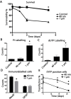
Results: Following the ex vivo extraction of adult mouse brains, mounted inside a medium-agarose matrix, we have exploited a precise slicing procedure using a custom built vibroslicer. To transfect these slices we used an improved biolistic transfection method using a custom made low-pressure barrel and novel DNA-coated nanoparticles (40 nm), which are drastically smaller than traditional microparticles. These nanoparticles also minimize tissue damage as seen by a significant reduction in lactate dehydrogenase activity as well as propidium iodide (PI) and dUTP labelling compared to larger traditional gold particles used on these OTBS. Furthermore, following EYFP exogene delivery by gene gun, the 40 nm treated OTBS displayed a significantly larger number of viable NeuN and EYFP positive cells. These OTBS expressed the exogenous proteins for many weeks.
Conclusions: Our described methodology of producing OTBS, which results in better reproducibility with less tissue damage, permits the exploitation of mature fully formed adult brains for advanced neurobiological studies. The novel 40 nm particles are ideal for the viable biolistic transfection of OTBS by reducing tissue stress while maintaining long term exogene expression.
Figures


Similar articles
-
Regioselective biolistic targeting in organotypic brain slices using a modified gene gun.J Vis Exp. 2014 Oct 24;(92):e52148. doi: 10.3791/52148. J Vis Exp. 2014. PMID: 25407047 Free PMC article.
-
Biolistic transfection of neuronal cultures using a hand-held gene gun.Nat Protoc. 2006;1(2):977-81. doi: 10.1038/nprot.2006.145. Nat Protoc. 2006. PMID: 17406333 Free PMC article.
-
Biolistic transfection and expression analysis of acute cortical slices.J Neurosci Methods. 2020 May 1;337:108666. doi: 10.1016/j.jneumeth.2020.108666. Epub 2020 Feb 28. J Neurosci Methods. 2020. PMID: 32119875 Free PMC article.
-
Nano-biolistics: a method of biolistic transfection of cells and tissues using a gene gun with novel nanometer-sized projectiles.BMC Biotechnol. 2011 Jun 10;11:66. doi: 10.1186/1472-6750-11-66. BMC Biotechnol. 2011. PMID: 21663596 Free PMC article.
-
Diolistics: incorporating fluorescent dyes into biological samples using a gene gun.Trends Biotechnol. 2007 Nov;25(11):530-4. doi: 10.1016/j.tibtech.2007.07.014. Epub 2007 Oct 22. Trends Biotechnol. 2007. PMID: 17945370 Free PMC article. Review.
Cited by
-
Correlated light and electron microscopy: ultrastructure lights up!Nat Methods. 2015 Jun;12(6):503-13. doi: 10.1038/nmeth.3400. Nat Methods. 2015. PMID: 26020503 Review.
-
Fluorescent labeling of dendritic spines in cell cultures with the carbocyanine dye "DiI".Front Neuroanat. 2014 May 9;8:30. doi: 10.3389/fnana.2014.00030. eCollection 2014. Front Neuroanat. 2014. PMID: 24847216 Free PMC article.
-
Nanobiolistics: An Emerging Genetic Transformation Approach.Methods Mol Biol. 2020;2124:141-159. doi: 10.1007/978-1-0716-0356-7_7. Methods Mol Biol. 2020. PMID: 32277452 Free PMC article.
-
A novel ex vivo Huntington's disease model for studying GABAergic neurons and cell grafts by laser microdissection.PLoS One. 2018 Mar 5;13(3):e0193409. doi: 10.1371/journal.pone.0193409. eCollection 2018. PLoS One. 2018. PMID: 29505597 Free PMC article.
-
Organotypic Cultures from the Adult CNS: A Novel Model to Study Demyelination and Remyelination Ex Vivo.Cell Mol Neurobiol. 2018 Jan;38(1):317-328. doi: 10.1007/s10571-017-0529-6. Epub 2017 Aug 9. Cell Mol Neurobiol. 2018. PMID: 28795301 Free PMC article. Review.
References
Publication types
MeSH terms
Substances
Grants and funding
LinkOut - more resources
Full Text Sources
Other Literature Sources
Research Materials
Miscellaneous

